- Gypsy Rose Blanchard separates from husband Ryan Anderson 3 months after her prison release: report New York Post
- Gypsy Rose Blanchard Announces Separation from Husband 3 Months After Prison Release PEOPLE
- Mom murderer Gypsy Rose Blanchard announces SPLIT from husband Ryan Scott Anderson just three months after rel Daily Mail
- Gypsy Rose Blanchard Splits from Husband Ryan Scott Anderson, 3 Months After Release From Prison Just Jared
- Gypsy Rose Blanchard and Husband Ryan Anderson Announce Separation 3 Months After Her Prison Release: Report Entertainment Tonight
Tag Archives: Separates
How the Brain Separates Perception From Memory
Summary: While previous studies point to an overlap between perception and memory, a new study finds the two are systematically different.
Source: NYU
The brain works in fundamentally different ways when remembering what we have seen compared to seeing something for the first time, a team of scientists has found.
While previous work had concluded there is significant overlap between these two processes, the new study, which appears in the journal Nature Communications, reveals they are systematically different.
“There are undoubtedly some similarities between the brain’s activity when people are seeing and remembering things, but there are also significant differences,” says Jonathan Winawer, a professor of psychology and neuroscience at New York University and the senior author of the paper.
“These distinctions are crucial to better understanding memory behavior and related afflictions.”
“We think these differences have to do with the architecture of the visual system itself and that the vision and memory processes produce different patterns of activity within this architecture,” adds Serra Favila, the paper’s lead author and an NYU doctoral student at the time of the study.
For decades, it was thought that recalling what we have seen—a sunset, a painting, another’s face—meant reactivating the same neuronal process used when seeing these images for the first time. However, the relationship between these activities—feedforward (vision) and feedback (memory)—is unclear.
To explore this, the research team, which also included Brice Kuhl, previously an assistant professor at NYU, conducted a series of experiments with human subjects.
Using functional MRI (fMRI) technology, the scientists measured the subjects’ visual cortex responses as they viewed images (simple geometric shapes in different locations on a computer screen) and, later, when they were asked to recall their make-up.
Varying the location of these visual shapes in the experiments allowed the researchers to monitor and understand memory activity in the visual system in a highly precise way.
The results showed some similarities between neuronal activity when initially processing these visual shapes and when asked to recall them—the parts of the visual cortex deployed when seeing something for the first time (perception) were also active during memory processing.
However, activity during memory also differed from activity during perception in highly systematic ways. Many of these differences stem from how visual scenes are mapped onto the brain. The brain has dozens of visual areas to process and store incoming images. These areas are arranged in a hierarchy—a long-understood characteristic.
More specifically, the primary visual cortex (V1) is at the bottom of the hierarchy because it is the first area to receive visual inputs, and it maps the visual scene in fine spatial detail. The signals are then passed along to subsequent brain maps for further processing—to the secondary visual cortex, or V2, and then V3, etc.
The initial processing by the primary visual cortex accurately captures the spatial arrangement of images while the higher brain areas, such as the secondary visual cortex, extract more complex information—What shape does an object have? What color is it? Is it a cup or bowl? But what is gained in complexity is lost in spatial precision.
“The tradeoff is that as these higher areas extract more complex information, they become less concerned about the exact spatial arrangement of the image,” explains Winawer.
In the Nature Communications study, the researchers found that during perception viewing a small object activated a small part of the primary visual cortex, a larger part of secondary visual cortex, and even larger parts of higher cortices.
This was expected due to the known properties of the visual hierarchy, they note. However, they found that this progression appears to be lost when recalling a visual stimulus (i.e., memory).
The scientists say this is akin to the way ink spreads on stacked pieces of paper. In perception, brain activity becomes more dispersed as you move up the organ’s hierarchy.
By contrast, in memory, the ink starts out at the top of the hierarchy, already dispersed, and cannot get narrower as it goes back down, thus the activity remains relatively constant.
See also


This loss of progression during memory may explain why remembering a scene is so different from seeing one, and why there tends to be so much less detail available in memory.
About this perception and memory research news
Author: Press Office
Source: NYU
Contact: Press Office – NYU
Image: The image is credited to Jonathan Winawer, NYU’s Department of Psychology/New York University
Original Research: Open access.
“Perception and memory have distinct spatial tuning properties in human visual cortex” by Serra E. Favila et al. Nature Communications
Abstract
Perception and memory have distinct spatial tuning properties in human visual cortex
Reactivation of earlier perceptual activity is thought to underlie long-term memory recall. Despite evidence for this view, it is unclear whether mnemonic activity exhibits the same tuning properties as feedforward perceptual activity.
Here, we leverage population receptive field models to parameterize fMRI activity in human visual cortex during spatial memory retrieval.
Though retinotopic organization is present during both perception and memory, large systematic differences in tuning are also evident. Whereas there is a three-fold decline in spatial precision from early to late visual areas during perception, this pattern is not observed during memory retrieval.
This difference cannot be explained by reduced signal-to-noise or poor performance on memory trials. Instead, by simulating top-down activity in a network model of cortex, we demonstrate that this property is well explained by the hierarchical structure of the visual system.
Together, modeling and empirical results suggest that computational constraints imposed by visual system architecture limit the fidelity of memory reactivation in sensory cortex.
Shanghai separates COVID-positive children from parents in virus fight
SHANGHAI, April 2 (Reuters) – Esther Zhao thought she was doing the right thing when she brought her 2-1/2-year-old daughter to a Shanghai hospital with a fever on March 26.
Three days later, Zhao was begging health authorities not to separate them after she and the little girl both tested positive for COVID-19, saying her daughter was too young to be taken away to a quarantine centre for children.
Doctors then threatened Zhao that her daughter would be left at the hospital, while she was sent to the centre, if she did not agree to transfer the girl to the Shanghai Public Health Clinical Center in the city’s Jinshan district.
Register now for FREE unlimited access to Reuters.com
Since her daughter was sent to the centre Zhao has had only one brief message that she was fine, sent through a group chat with doctors, despite repeated pleas for information from Zhao and her husband, who is in a separate quarantine site after also testing positive.
“There have been no photos at all… I’m so anxious, I have no idea what situation my daughter is in,” she said on Saturday through tears, still stuck at the hospital she went to last week. “The doctor said Shanghai rules is that children must be sent to designated points, adults to quarantine centres and you’re not allowed to accompany the children.”
Zhao is panicking even more after images of crying children at a Shanghai health facility went viral in China. The anonymous poster said these were children who had tested positive for COVID-19 and been separated from their parents at the Jinshan centre.
The photos and videos posted on China’s Weibo and Douyin social media platforms showed wailing babies kept three to a cot. In one video, a groaning toddler crawls out of a room with four child-sized beds pushed against the wall. While a few adults can be seen in the videos, they are outnumbered by the number of children.
Reuters could not immediately verify the images, but a source familiar with the facility confirmed they were taken at the Jinshan facility.
The Shanghai Public Health Clinical Center said, however, that the photos and videos circulating on internet were not of a “Jinshan infant quarantine facility” but were scenes taken when the hospital was moving its paediatric ward to another building to cope with a rising number of COVID paediatric patients.
This was done to “improve the hospital environment”, it said on its official WeChat account on Saturday, adding that it had organised for more pediatric workers and would strengthen communication with the children’s parents.
“Paediatric patients admitted to our hospital… are guaranteed medical treatment and their daily needs taken care of,” it said.
Later on Saturday the Shanghai rumour buster WeChat account, which is backed by China’s cyberspace watchdog, published four photos that it said showed the children’s current situation at the Jinshan centre.
One of the photos showed young children sitting in and standing around beds that were arranged neatly in two rows, though no adults were pictured. In another photo, a hazmat-suited person attends to a baby lying in a cot. Only one other adult, also in a hazmat suit, can be seen in the two other photos.
The Shanghai government referred Reuters to the hospital’s statement and declined to comment further.
A Shanghai health official said last week that hospitals that were treating COVID-positive children maintained online communications with their parents.
POST DELETED
By Saturday, the original post had been deleted from Weibo, but thousands of people continued to comment and repost the images. “This is horrific,” said one. “How could the government come up with such a plan?,” said another.
In some cases children as young as 3 months old are being separated from their breastfeeding mothers, according to posts in a quarantine hospital WeChat group shared with Reuters. In one room described in a post, there are eight children without an adult.
In another case, more than 20 children from a Shanghai kindergarten aged 5 to 6 were sent to a quarantine centre without their parents, a source familiar with the situation said.
Since Shanghai’s latest outbreak began about a month ago, authorities have locked down its 26 million people in a two-stage process that began on Monday.
While the number of cases in Shanghai is small by global standards, Chinese authorities have vowed to stick with “dynamic clearance”, aiming to test for, trace and centrally quarantine all positive cases.
The U.S., French and Italian foreign consulates have warned their citizens in Shanghai that family separations could happen as Chinese authorities executed COVID curbs, according to notices seen by Reuters.
Shanghai on Saturday reported 6,051 locally transmitted asymptomatic COVID-19 cases and 260 symptomatic cases for April 1, versus 4,144 asymptomatic cases and 358 symptomatic ones on the previous day.
Register now for FREE unlimited access to Reuters.com
Reporting by Brenda Goh and Engen Tham, Additional reporting by Winni Zhou; Editing by Christian Schmollinger, William Mallard and Clelia Oziel
Our Standards: The Thomson Reuters Trust Principles.
Researchers Discover How the Human Brain Separates, Stores, and Retrieves Memories
NIH-funded study identifies brain cells that form boundaries between discrete events.
Researchers have identified two types of cells in our brains that are involved in organizing discrete memories based on when they occurred. This finding improves our understanding of how the human brain forms memories and could have implications in memory disorders such as 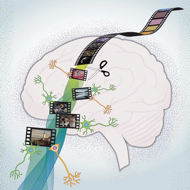

Researchers recorded the brain activity of participants as they watched videos, and they noticed two distinct groups of cells that responded to different types of boundaries by increasing activity. Credit: Rutishauser lab, Cedars-Sinai Medical Center
To study this, Rutishauser and his colleagues worked with 20 patients who were undergoing intracranial recording of brain activity to guide surgery for treatment of their drug-resistant epilepsy. They looked at how the patients’ brain activity was affected when shown film clips containing different types of “cognitive boundaries”—transitions thought to trigger changes in how a memory is stored and that mark the beginning and end of memory “files” in the brain.
The first type, referred to as a “soft boundary,” is a video containing a scene that then cuts to another scene that continues the same story. For example, a baseball game showing a pitch is thrown and, when the batter hits the ball, the camera cuts to a shot of the fielder making a play. In contrast, a “hard boundary” is a cut to a completely different story—imagine if the batted ball were immediately followed by a cut to a commercial.
Jie Zheng, Ph.D., postdoctoral fellow at Children’s Hospital Boston and first author of the study, explained the key difference between the two boundaries.
“Is this a new scene within the same story, or are we watching a completely different story? How much the narrative changes from one clip to the next determines the type of cognitive boundary,” said Zheng.
The researchers recorded the brain activity of participants as they watched the videos, and they noticed two distinct groups of cells that responded to different types of boundaries by increasing their activity. One group, called “boundary cells” became more active in response to either a soft or hard boundary. A second group, referred to as “event cells” responded only to hard boundaries. This led to the theory that the creation of a new memory occurs when there is a peak in the activity of both boundary and event cells, which is something that only occurs following a hard boundary.
One analogy to how memories might be stored and accessed in the brain is how photos are stored on your phone or computer. Often, photos are automatically grouped into events based on when and where they were taken and then later displayed to you as a key photo from that event. When you tap or click on that photo, you can drill down into that specific event.
“A boundary response can be thought of like creating a new photo event,” said Dr. Rutishauser. “As you build the memory, it’s like new photos are being added to that event. When a hard boundary occurs, that event is closed and a new one begins. Soft boundaries can be thought of to represent new images created within a single event.”
The researchers next looked at memory retrieval and how this process relates to the firing of boundary and event cells. They theorized that the brain uses boundary peaks as markers for “skimming” over past memories, much in the way the key photos are used to identify events. When the brain finds a firing pattern that looks familiar, it “opens” that event.
Two different memory tests designed to study this theory were used. In the first, the participants were shown a series of still images and were asked whether they were from a scene in the film clips they just watched. Study participants were more likely to remember images that occurred soon after a hard or soft boundary, which is when a new “photo” or “event” would have been created.
The second test involved showing pairs of images taken from film clips that they had just watched. The participants were then asked which of the two images had appeared first. It turned out that they had a much harder time choosing the correct image if the two occurred on different sides of a hard boundary, possibly because they had been placed in different “events.”
These findings provide a look into how the human brain creates, stores, and accesses memories. Because event segmentation is a process that can be affected in people living with memory disorders, these insights could be applied to the development of new therapies.
In the future, Dr. Rutishauser and his team plan to look at two possible avenues to develop therapies related to these findings. First, neurons that use the chemical dopamine, which are most-known for their role in reward mechanisms, may be activated by boundary and event cells, suggesting a possible target to help strengthen the formation of memories.
Second, one of the brain’s normal internal rhythms, known as the theta rhythm, has been connected to learning and memory. If event cells fired in time with that rhythm, the participants had an easier time remembering the order of the images that they were shown. Because deep brain stimulation can affect theta rhythms, this could be another avenue for treating patients with certain memory disorders.
Reference: “Neurons detect cognitive boundaries to structure episodic memories in humans” by Jie Zheng, Andrea G. P. Schjetnan, Mar Yebra, Bernard A. Gomes, Clayton P. Mosher, Suneil K. Kalia, Taufik A. Valiante, Adam N. Mamelak, Gabriel Kreiman and Ueli Rutishauser, 7 March 2022, Nature Neuroscience.
DOI: 10.1038/s41593-022-01020-w
This project was made possible by a multi-institutional consortium through the NIH BRAIN Initiative’s Research on Humans program. Institutions involved in this study were Cedars-Sinai Medical Center, Children’s Hospital Boston (site PI Gabriel Kreiman, Ph.D.), and Toronto Western Hospital (site PI Taufik Valiante, M.D., Ph.D.). The study was funded by the NIH BRAIN Initiative (NS103792, NS117839), the National Science Foundation, and Brain Canada.
The BRAIN Initiative® is a registered trademark of the U.S. Department of Health and Human Services.
The NIH BRAIN Initiative is managed by 10 institutes whose missions and current research portfolios complement the goals of The BRAIN Initiative®: National Center for Complementary and Integrative Health, National Eye Institute, National Institute on Aging, National Institute on Alcohol Abuse and Alcoholism, National Institute of Biomedical Imaging and Bioengineering, Eunice Kennedy Shriver National Institute of Child Health and Human Development, National Institute on Drug Abuse, National Institute on Deafness and other Communication Disorders, National Institute of Mental Health, and National Institute of Neurological Disorders and Stroke.
NINDS is the nation’s leading funder of research on the brain and nervous system. The mission of NINDS is to seek fundamental knowledge about the brain and nervous system and to use that knowledge to reduce the burden of neurological disease.
About the National Institutes of Health (NIH): NIH, the nation’s medical research agency, includes 27 Institutes and Centers and is a component of the U.S. Department of Health and Human Services. NIH is the primary federal agency conducting and supporting basic, clinical, and translational medical research, and is investigating the causes, treatments, and cures for both common and rare diseases.