- Precision functional MRI mapping reveals distinct connectivity patterns for depression associated with traumatic brain injury Science
- Redefining Depression: TBI Affective Syndrome Discovered Neuroscience News
- Depression after a brain injury is a distinct condition, study finds. That could change how it’s treated. AOL
- Why depression after traumatic brain injury is distinct — and less likely to respond to standard treatment STAT
- Abnormal Brain Folding a Biomarker for Major Depressive Disorder Neuroscience News
- View Full Coverage on Google News
Tag Archives: Mapping
Most Detailed Mapping of Our Brains’ Memory Bank Reveals Something Surprising : ScienceAlert
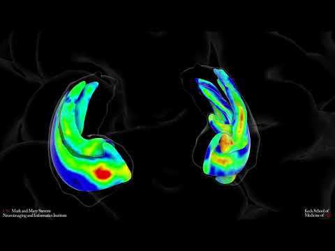
Scientists have created the most detailed map yet of the neural highways connecting our gray matter’s memory bank – the hippocampus – with the rest of the brain, revealing unexpected patterns of connections between regions.
“We were surprised to find fewer connections between the hippocampus and frontal cortical areas and more connections with early visual processing areas than we expected to see,” says University of Sydney psychologist Marshall Dalton.
While there’s still a lot of debate over the precise role of the hippocampus in memory, neurologists are confident it plays a key part in constructing memory and integrating it with our perception to allow us to make decisions about the future.
A better understanding of how the hippocampus works in context with other areas of the brain could one day help us tackle memory decline.
Using a new imaging technique called diffusion weighted imaging – a type of MRI scan that uses diffusion of water molecules through tissues to generate contrast – Dalton and colleagues created a high-resolution map of the connections between the hippocampus and cerebral cortex from the brains of seven adult females under 35 years old.
“We have now developed a tailored method that allows us to confirm where within the hippocampus different cortical areas are connecting. And that hasn’t been done before in a living human brain,” says Dalton.
“What we’ve done is take a much more detailed look at the white matter pathways, which are essentially the highways of communication between different areas of the brain.”
The researchers found the hippocampus has different messaging networks, each linked to specific areas of the cortex. Our previous knowledge of these connections stems from dissections of primate brains, and the resulting brain map largely aligned with these.
But the researchers discovered a much higher level of connections in the visual processing area of the human brain and fewer in the frontal cortical areas.
The post-mortem analysis carried out on non-human primates can detect finer details down to a cellular level, so it may just be that we haven’t been able to resolve all those connections in humans yet.
“Or it could be that the human hippocampus really does have a smaller number of connections with frontal areas than we expect and greater connectivity with visual areas of the brain,” Dalton explains.
“This makes sense considering the hippocampus plays an important role not only in memory but also imagination and our ability to construct mental images in our mind’s eye.”
Other recent studies have also found associations between these areas of the brain. The team is curious to see if similar patterns are consistent across humans of different demographics.
“As the neocortex expanded, perhaps humans evolved different patterns of connectivity to facilitate human-specific memory and visualization functions which, in turn, may underpin human creativity,” Dalton continues.
“It’s a bit of a puzzle – we just don’t know. But we love puzzles and will keep investigating.”
This research was published in eLife.
New mapping method could aid exploration of moon, Mars and beyond
Researchers have discovered a method for making high-resolution maps of planetary surfaces like the moon’s by combining available imagery and topography data.
Mapping the complex and diverse surface of a world like the moon in detailed resolution is challenging because laser altimeters, which measure changes in altitudes, operate at much lower resolution than cameras. And although photographs offer a sense of surface features, it’s difficult to translate images into specific heights and depths.
However, a new method combines topographic data with degrees of shading of sunlight present in optical images to provide much more accurate assessments of landforms and structures. The development could help both astronauts and robotic explorers in the future.
Related: Rogue rocket’s moon crash site spotted by NASA probe (photos)
The new method was developed by Iris Fernandes, a now former Ph.D. student at the Niels Bohr Institute of the University of Copenhagen in Denmark.
Different missions deliver very different data resolutions, so combining different sources of data in different resolutions presents a huge mathematical problem. What Fernandes and her then-supervisor, geophysicist Klaus Mosegaard, did differently was to focus on the mathematics of the issue, narrowing down the challenge to determine whether an equation could solve the problem.
“And it did,” Fernandes said in a statement (opens in new tab), explaining that they had found a “mathematical key to a door that had remained closed for many years.”
The new approach also requires much less computational power and is much faster than previous methods, according to the researchers.
The researchers demonstrated the technique on the moon. By combining high-resolution images from NASA’s Lunar Reconnaissance Orbiter‘s LROC camera with lower-resolution data from the spacecraft’s Lunar Orbiter Laser Altimeter (LOLA), the authors of the paper state they increased the resolution of the topography from around 200 feet (60 meters) per pixel to 2.95 feet (0.9 m) per pixel.
Going forward, the authors argue, the method can be applied to extract precise topographic details on rock formations on planetary bodies such as the moon, Mars, asteroids and any other worlds for which topographic data is available.
The method can support a range of different purposes, including determining the safety of astronauts and rovers or finding geologically interesting sites, and it can be applied to images coming from satellites, rovers or other spacecraft.
The research is described in an article published June 8 in the journal Planetary and Space Science (opens in new tab).
Follow us on Twitter @Spacedotcom (opens in new tab) and on Facebook (opens in new tab).
HDMI 2.1a to get new standards for next-gen HDMI displays, including Source-Based Tone Mapping
We recently reported that the HDMI Licensing Administrator stopped labeling newer HDMI displays with the 2.0 version, and allowed manufacturers the ability to utilize the HDMI 2.1 moniker, as long as it fits the standards already set. This potentially can cause a manufacturer to label an HDMI 2.0 display as the 2.1 version to falsify claims to consumers and was seen by a Chinese seller practicing that exact scenario.
Now, there is a new HDMI 2.1 variant about to be released at some point in the future with the moniker of “HDMI 2.1a.”
The HDMI Licensing Administrator confirms new standards for HDMI 2.1 with the addition of SBTM
What is currently odd is the information about HDMI 2.1a, as it will be soon called, was appearing on the Licensing Administration’s page but has since mysteriously disappeared with no explanations. It is possible that there are a few last-minute decisions to finalize the new standard for use, but that in and of itself is pure speculation. VideoCardz has mentioned that the information can be found via Google Cache. One of the newest additions to the HDMI 2.1a is the use of Source-Based Tone Mapping or SBTM.
Source-Based Tone Mapping (SBTM) is a new HDR feature that allows a portion of the HDR mapping to be performed by the Source device instead of the Display device. SBTM is especially useful in cases where HDR and SDR video or graphics are combined together into a single picture, such as picture-in-picture or a program guide with an integrated video window. SBTM also enables PCs and gaming devices to automatically produce an optimized HDR signal in order to maximize the utilization of the display’s HDR capabilities without manual user configuration of the Source device.
— HDMI.org


Gamers and PC enthusiasts will see the most use of the new technology, due to the ability for multiwindowed environments. The source device, in essence, controls the tone mapping. This process allows separate windows to display either SDR or HDR optimized content simultaneously. TFTCentral states that the new SBTM is not a replacement for the current HDR technology (HDR10 and HLG, as well as other offerings). SBTM is set to deepen the HDR experience by offering more capability for optimization with HDR-capable devices.
Source: TFTCentral, VideoCardz
Why Scientists Have Spent Years Mapping This Creature’s Brain
The brain of a fruit fly is the size of a poppy seed and about as easy to overlook.
“Most people, I think, don’t even think of the fly as having a brain,” said Vivek Jayaraman, a neuroscientist at the Janelia Research Campus of the Howard Hughes Medical Institute in Virginia. “But, of course, flies lead quite rich lives.”
Flies are capable of sophisticated behaviors, including navigating diverse landscapes, tussling with rivals and serenading potential mates. And their speck-size brains are tremendously complex, containing some 100,000 neurons and tens of millions of connections, or synapses, between them.
Since 2014, a team of scientists at Janelia, in collaboration with researchers at Google, have been mapping these neurons and synapses in an effort to create a comprehensive wiring diagram, also known as a connectome, of the fruit fly brain.
The work, which is continuing, is time-consuming and expensive, even with the help of state-of-the-art machine-learning algorithms. But the data they have released so far is stunning in its detail, composing an atlas of tens of thousands of gnarled neurons in many crucial areas of the fly brain.
And now, in an enormous new paper, being published on Tuesday in the journal eLife, neuroscientists are beginning to show what they can do with it.
By analyzing the connectome of just a small part of the fly brain — the central complex, which plays an important role in navigation — Dr. Jayaraman and his colleagues identified dozens of new neuron types and pinpointed neural circuits that appear to help flies make their way through the world. The work could ultimately help provide insight into how all kinds of animal brains, including our own, process a flood of sensory information and translate it into appropriate action.
It is also a proof of principle for the young field of modern connectomics, which was built on the promise that constructing detailed diagrams of the brain’s wiring would pay scientific dividends.
“It’s really extraordinary,” Dr. Clay Reid, a senior investigator at the Allen Institute for Brain Science in Seattle, said of the new paper. “I think anyone who looks at it will say connectomics is a tool that we need in neuroscience — full stop.”
‘Your fly brain is cooked’
The only complete connectome in the animal kingdom belongs to the humble roundworm, C. elegans. The pioneering biologist Sydney Brenner, who would later go on to win a Nobel Prize, started the project in the 1960s. His small team spent years on it, using colored pens to trace all 302 neurons by hand.
“Brenner realized that to understand the nervous system you had to know its structure,” said Scott Emmons, a neuroscientist and geneticist at Albert Einstein College of Medicine, who later used digital techniques to create new C. elegans connectomes. “And that’s true across biology. Structure is so important.”
Brenner and his colleagues published their landmark paper, which clocked in at 340 pages, in 1986.
But the field of modern connectomics did not take off until the 2000s, when advances in imaging and computing finally made it feasible to map the connections in larger brains. In recent years, research teams around the world have started assembling connectomes of zebrafish, songbirds, mice, humans and more.
When the Janelia Research Campus opened in 2006, Gerald Rubin, its founding director, set his sights on the fruit fly. “I don’t want to offend any of my worm colleagues, but I think flies are the simplest brain that actually does interesting, complex behavior,” Dr. Rubin said.
Several different teams at Janelia have embarked on fly connectome projects in the years since, but the work that led to the new paper began in 2014, with the brain of a single, five-day-old female fruit fly.
Researchers cut the fly brain into slabs and then used a technique known as focused-ion beam scanning electron microscopy to image them, layer by painstaking layer. The microscope essentially functioned like a very tiny, very precise nail file, filing away an exceedingly thin layer of the brain, snapping a picture of the exposed tissue and then repeating the process until nothing remained.
“You’re simultaneously imaging and cutting off little slices of the fly brain, so they don’t exist after you’re done,” Dr. Jayaraman said. “So if you screw something up, you’re done. Your goose is cooked — or your fly brain is cooked.”
The team then used computer vision software to stitch the millions of resulting images back together into a single, three-dimensional volume and sent it off to Google. There, researchers used advanced machine-learning algorithms to identify each individual neuron and trace its twisting branches.
Finally, the Janelia team used additional computational tools to pinpoint the synapses, and human researchers proofread the computers’ work, correcting errors and refining the wiring diagrams.
Last year, the researchers published the connectome for what they called the “hemibrain,” a large portion of the central fly brain, which includes regions and structures that are crucial for sleep, learning and navigation.
The connectome, which is accessible free online, includes about 25,000 neurons and 20 million synapses, far more than the C. elegans connectome.
“It’s a dramatic scaling up,” said Cori Bargmann, a neuroscientist at the Rockefeller University in New York. “This is a tremendous step toward the goal of working out the connectivity of the brain.”
Welcome to orientation
Once the hemibrain connectome was ready, Dr. Jayaraman, an expert on the neuroscience of fly navigation, was eager to dive into the data on the central complex.
The brain region, which contains nearly 3,000 neurons and is present in all insects, helps flies build an internal model of their spatial relationship to the world and then select and execute behaviors appropriate for their circumstances, such as searching for food when they are hungry.
“You’re telling me you can give me the wiring diagram for something like this?” Dr. Jayaraman said. “This is better industrial espionage than you could get by getting insights into the Apple iPhone.”
He and his colleagues pored over the connectome data, studying how the region’s neural circuits were put together.
For instance, Hannah Haberkern, a postdoctoral associate in Dr. Jayaraman’s lab, analyzed the neurons that send sensory information to the ellipsoid body, a doughnut-shape structure that acts as the fly’s internal compass.
Dr. Haberkern found that neurons that are known to transmit information about the polarization of light — a global environmental cue that many animals use for navigation — made more connections to the compass neurons than did neurons that transmit information about other visual features and landmarks.
The neurons dedicated to polarization of light also connect to — and are capable of strongly inhibiting — brain cells that provide information about other navigational cues.
The researchers hypothesize that fly brains may be wired to prioritize information about the global environment when they are navigating — but also that these circuits are flexible, so that when such information is inadequate, they can pay more attention to local features of the landscape. “They have all these fallback strategies,” Dr. Haberkern said.
Fruit fly phone home
Other members of the research team identified specific neural pathways that seem well suited to helping the fly keep track of its head and body orientation, anticipate its future orientation and traveling direction, calculate its current orientation relative to another desired location and then move in that direction.
Imagine, for instance, that a hungry fly temporarily abandons a rotting banana to see whether it can rustle up something better. But after a (literally) fruitless few minutes of exploration, it wants to return to its previous meal.
The connectome data suggests that certain brain cells, technically known as PFL3 neurons, help the fly pull off this maneuver. These neurons receive two critical inputs: They get signals from neurons that track the direction the fly is facing as well as from neurons that may be keeping tabs on the direction of the banana.
After receiving those signals, the PFL3 neurons then send out their own message to a set of turning neurons that prompt the fly to veer off in the correct direction. Dinner is served, again.
“Being able to trace that activity through that circuit — from sensory back to motor through this complex intermediate circuit — is really amazing,” said Brad Hulse, a research scientist in Dr. Jayaraman’s lab who led this part of the analysis. The connectome, he added, “showed us a lot more than we thought it was going to.”
And the group’s paper — a draft of which includes 75 figures and stretches to 360 pages — is just the beginning.
“It just really provides this ground truth for exploring this brain region further,” said Stanley Heinze, an expert on insect neuroscience at Lund University in Sweden. “It’s just enormously impressive.”
And just plain enormous. “I wouldn’t really treat it as a paper but more as a book,” Dr. Heinze said.
In fact, the paper is so large that the preprint server bioRxiv initially declined to publish it, perhaps because the administrators — understandably — thought it actually was a book, Dr. Jayaraman said. (The server ultimately did post the study, after a few extra days of processing, he noted.)
The paper’s publication in the journal eLife “required some special permissions and back-and-forth with editorial staff,” Dr. Jayaraman added.
Fly-ing lessons
There are limitations to what a snapshot of a single brain at a single moment in time can reveal, and connectomes do not capture everything of interest in an animal brain. (Janelia’s hemibrain connectome omits glial cells, for instance, which perform all sorts of important tasks in the brain.)
Dr. Jayaraman and his colleagues stressed that they would not have been able to infer so much from the connectome if not for decades of prior research, by many other scientists, into fruit fly behavior and basic neuron physiology and function, as well as theoretical neuroscience work.
But the wiring diagrams can help researchers investigate existing theories and generate better hypotheses, figuring out what questions to ask and which experiments to conduct.
“Now what we’re really excited about is taking those ideas that the connectome inspired and going back to the microscope, going back to our electrodes and actually recording the brain and seeing if those ideas are true,” Dr. Hulse said.
Of course, one could — and some have — asked why a fruit fly’s brain circuitry matters.
“I get asked this at the holidays a lot,” Dr. Hulse said.
Flies are not mice or chimps or humans, but their brains perform some of the same basic tasks. Understanding the basic neural circuitry in an insect could provide important clues to how other animal brains approach similar problems, said David Van Essen, a neuroscientist at Washington University in St. Louis.
Gaining a deep understanding of the fly’s brain “also gives us insights that are very relevant to the understanding of mammalian, and even human, brains and behavior,” he said.
Creating connectomes of larger, more complex brains will be enormously challenging. The mouse brain contains roughly 70 million neurons, the human brain a whopping 86 billion.
But the central complex paper is decidedly not a one-off; detailed studies of regional mouse and human connectomes are currently in the pipeline, Dr. Reid said: “There’s a lot more to come.”
Journal editors, consider yourselves warned.
Mapping the Universe’s Earliest Structures and Dark Matter Distribution With COSMOS-Webb
The COSMOS-Webb survey will map 0.6 square degrees of the sky—about the area of three full Moons—using the James Webb Space Telescope’s Near Infrared Camera (NIRCam) instrument, while simultaneously mapping a smaller 0.2 square degrees with the Mid Infrared Instrument (MIRI). The jagged edges of the Hubble field’s outline are due to the separate images that make up the survey field. Credit: Jeyhan Kartaltepe (RIT); Caitlin Casey (UT Austin); and Anton Koekemoer (STScI) Graphic Design Credit: Alyssa Pagan (STScI)
This ambitious program will study half a million galaxies in a field the size of three full Moons.
Peering deeply into a huge patch of sky the size of three full Moons, 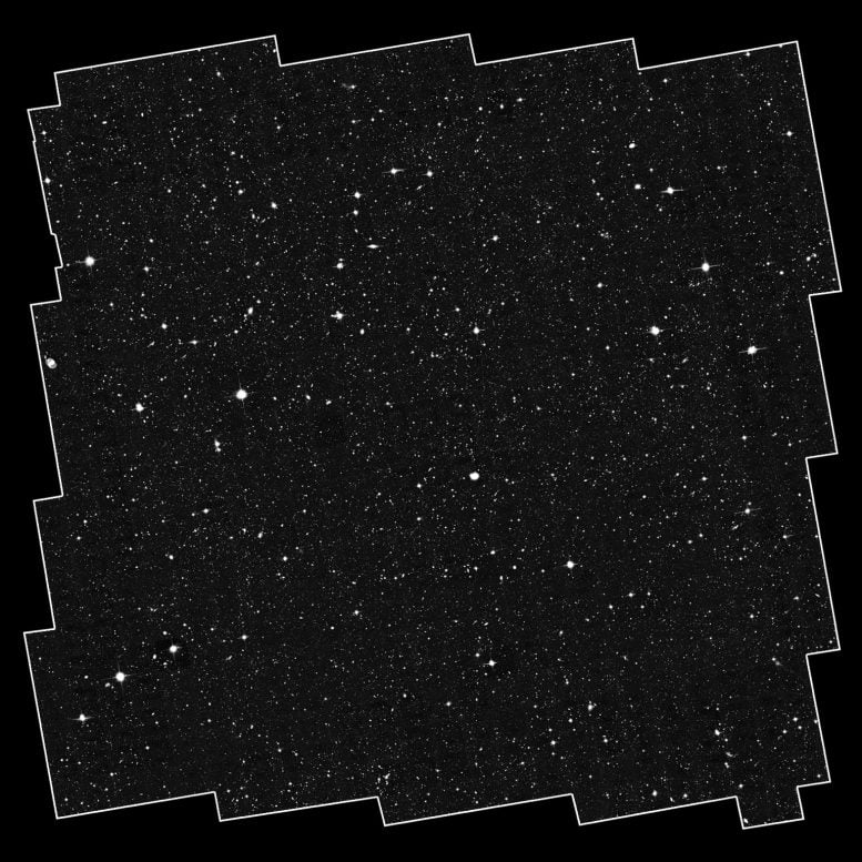

This sea of galaxies is the complete, original COSMOS field from the Hubble Space Telescope’s Advanced Camera for Surveys (ACS). The full mosaic is a composite of 575 separate ACS images, where each ACS image is about one-tenth the diameter of the full Moon. The jagged edges of the outline are due to the separate images that make up the survey field. Credit: Anton Koekemoer (STScI) and Nick Scoville (Caltech)
When NASA’s James Webb Space Telescope begins science operations in 2022, one of its first tasks will be an ambitious program to map the earliest structures in the universe. Called COSMOS-Webb, this wide and deep survey of half-a-million galaxies is the largest project Webb will undertake during its first year.
With more than 200 hours of observing time, COSMOS-Webb will survey a large patch of the sky—0.6 square degrees—with the Near-Infrared Camera (NIRCam). That’s the size of three full moons. It will simultaneously map a smaller area with the Mid-Infrared Instrument (MIRI).

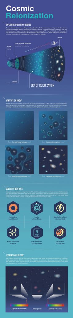
More than 13 billion years ago, during the Era of Reionization, the universe was a very different place. The gas between galaxies was largely opaque to energetic light, making it difficult to observe young galaxies. What allowed the universe to become completely ionized, or transparent, eventually leading to the “clear” conditions detected in much of the universe today? The James Webb Space Telescope will peer deep into space to gather more information about objects that existed during the Era of Reionization to help us understand this major transition in the history of the universe. Credit: NASA, ESA, and J. Kang (STScI)
It’s a large chunk of sky, which is pretty unique to the COSMOS-Webb program. Most Webb programs are drilling very deep, like pencil-beam surveys that are studying tiny patches of sky,” explained Caitlin Casey, an assistant professor at the University of Texas at Austin and co-leader of the COSMOS-Webb program. “Because we’re covering such a large area, we can look at large-scale structures at the dawn of galaxy formation. We will also look for some of the rarest galaxies that existed early on, as well as map the large-scale dark matter distribution of galaxies out to very early times.”
(Dark matter does not absorb, reflect, or emit light, so it cannot be seen directly. We know that dark matter exists because of the effect it has on objects that we can observe.)
COSMOS-Webb will study half-a-million galaxies with multi-band, high-resolution, near-infrared imaging, and an unprecedented 32,000 galaxies in the mid-infrared. With its rapid public release of the data, this survey will be a primary legacy dataset from Webb for scientists worldwide studying galaxies beyond the Milky Way.
Building on Hubble’s Achievements
The COSMOS survey began in 2002 as a Hubble program to image a much larger patch of sky, about the area of 10 full moons. From there, the collaboration snowballed to include most of the world’s major telescopes on Earth and in space. Now COSMOS is a multi-wavelength survey that covers the entire spectrum from the X-ray through the radio.
Because of its location on the sky, the COSMOS field is accessible to observatories around the world. Located on the celestial equator, it can be studied from both the northern and southern hemispheres, resulting in a rich and diverse treasury of data.
“COSMOS has become the survey that a lot of extragalactic scientists go to in order to conduct their analyses because the data products are so widely available, and because it covers such a wide area of the sky,” said Rochester Institute of Technology’s Jeyhan Kartaltepe, assistant professor of physics and co-leader of the COSMOS-Webb program. “COSMOS-Webb is the next installment of that, where we’re using Webb to extend our coverage in the near- and mid-infrared part of the spectrum, and therefore pushing out our horizon, how far away we’re able to see.”
The ambitious COSMOS-Webb program will build upon previous discoveries to make advances in three particular areas of study, including: revolutionizing our understanding of the Reionization Era; looking for early, fully evolved galaxies; and learning how dark matter evolved with galaxies’ stellar content.
Goal 1: Revolutionizing Our Understanding of the Reionization Era
Soon after the big bang, the universe was completely dark. Stars and galaxies, which bathe the cosmos in light, had not yet formed. Instead, the universe consisted of a primordial soup of neutral hydrogen and helium atoms and invisible dark matter. This is called the cosmic dark ages.
After several hundred million years, the first stars and galaxies emerged and provided energy to reionize the early universe. This energy ripped apart the hydrogen atoms that filled the universe, giving them an electric charge and ending the cosmic dark ages. This new era where the universe was flooded with light is called the Reionization Era.
The first goal of COSMOS-Webb focuses on this epoch of reionization, which took place from 400,000 to 1 billion years after the big bang. Reionization likely happened in little pockets, not all at once. COSMOS-Webb will look for bubbles showing where the first pockets of the early universe were reionized. The team aims to map the scale of these reionization bubbles.
“Hubble has done a great job of finding handfuls of these galaxies out to early times, but we need thousands more galaxies to understand the reionization process,” explained Casey.
Scientists don’t even know what kind of galaxies ushered in the Reionization Era, whether they’re very massive or relatively low-mass systems. COSMOS-Webb will have a unique ability to find very massive, rare galaxies and see what their distribution is like in large-scale structures. So, are the galaxies responsible for reionization living in the equivalent of a cosmic metropolis, or are they mostly evenly distributed across space? Only a survey the size of COSMOS-Webb can help scientists to answer this.
Goal 2: Looking for Early, Fully Evolved Galaxies
COSMOS-Webb will search for very early, fully evolved galaxies that shut down star birth in the first 2 billion years after the big bang. Hubble has found a handful of these galaxies, which challenge existing models about how the universe formed. Scientists struggle to explain how these galaxies could have old stars and not be forming any new stars so early in the history of the universe.
With a large survey like COSMOS-Webb, the team will find many of these rare galaxies. They plan detailed studies of these galaxies to understand how they could have evolved so rapidly and turned off star formation so early.
Goal 3: Learning How Dark Matter Evolved with Galaxies’ Stellar Content
COSMOS-Webb will give scientists insight into how dark matter in galaxies has evolved with the galaxies’ stellar content over the universe’s lifetime.
Galaxies are made of two types of matter: normal, luminous matter that we see in stars and other objects, and invisible dark matter, which is often more massive than the galaxy and can surround it in an extended halo. Those two kinds of matter are intertwined in galaxy formation and evolution. However, presently there’s not much knowledge about how the dark matter mass in the halos of galaxies formed, and how that dark matter impacts the formation of the galaxies.
COSMOS-Webb will shed light on this process by allowing scientists to directly measure these dark matter halos through “weak lensing.” The gravity from any type of mass—whether it’s dark or luminous—can serve as a lens to “bend” the light we see from more distant galaxies. Weak lensing distorts the apparent shape of background galaxies, so when a halo is located in front of other galaxies, scientists can directly measure the mass of the halo’s dark matter.
“For the first time, we’ll be able to measure the relationship between the dark matter mass and the luminous mass of galaxies back to the first 2 billion years of cosmic time,” said team member Anton Koekemoer, a research astronomer at the Space Telescope Science Institute in Baltimore, who helped design the program’s observing strategy and is in charge of constructing all the images from the program. “That’s a crucial epoch for us to try to understand how the galaxies’ mass was first put in place, and how that’s driven by the dark matter halos. And that can then feed indirectly into our understanding of galaxy formation.”
Quickly Sharing Data with the Community
COSMOS-Webb is a Treasury program, which by definition is designed to create datasets of lasting scientific value. Treasury Programs strive to solve multiple scientific problems with a single, coherent dataset. Data taken under a Treasury Program usually has no exclusive access period, enabling immediate analysis by other researchers.
“As a Treasury Program, you are committing to quickly releasing your data and your data products to the community,” explained Kartaltepe. “We’re going to produce this community resource and make it publicly available so that the rest of the community can use it in their scientific analyses.”
Koekemoer added, “A Treasury Program commits to making publicly available all these science products so that anyone in the community, even at very small institutions, can have the same, equal access to the data products and then just do the science.”
COSMOS-Webb is a Cycle 1 General Observers program. General Observers programs were competitively selected using a dual-anonymous review system, the same system that is used to allocate time on Hubble.
The James Webb Space Telescope will be the world’s premier space science observatory when it launches in 2021. Webb will solve mysteries in our solar system, look beyond to distant worlds around other stars, and probe the mysterious structures and origins of our universe and our place in it. Webb is an international program led by NASA with its partners, ESA (European Space Agency) and the Canadian Space Agency.
Mapping the ice on Mars that could support future missions
Enlarge / While we know of locations with ice on Mars, not all of them are in places we’d want to land.
Over the past couple decades, plans to go to Mars or return to the Moon for longer stays have gradually moved away from sci-fi tinged “what if” scenarios and shifted to something that resembles actual planning. And those plans invariably include extracting water from local ice deposits. This water would help support any astronauts during their stay, cutting down on the weight we’d have to shift out of Earth orbit. But it could also be a source of hydrogen that helps power the astronaut’s return trip to Earth.
That obviously means we want to land where the water is. On the Moon, this has meant focusing on the lunar poles, where deep craters create permanent shadows that can hold ice at temperatures where it’s stable. On Mars, the situation is considerably more complicated. In response to some NASA pilot funding, a team of scientists set up the SWIM projectM, for Subsurface Water Ice Mapping on Mars, to analyze the data. The project has now published a progress report showing a lot of ice deposits in areas we might want to land.
No poles, please
Whether or not water ice is stable on the Moon is determined entirely by sunlight exposure. As long as the Sun is never visible in a location, ice can survive. Mars is substantially more complicated, with an atmosphere that distributes heat and makes the temperature extremes far more moderate, plus orbital wobbles that ensure seasonal changes in temperature.
Mars does have polar ice, but the number of these deposits changes with the seasons (and a lot of it is frozen carbon dioxide). Further from the poles, there’s a region where temperatures would allow water ice to be stable, should it form there. But that region’s still far from the equator, which means more extreme cold and less solar energy for any photovoltaic equipment we might bring with us. Ideally, it would be nice to find some ice in temperate regions, and some reports have suggested locations where it might reside.
The SWIM team decided to take a far more comprehensive approach, using data from multiple instruments to try to establish a degree of confidence in the presence of water. To do so, the team developed its own ice scoring system.
That data comes from a number of instruments we’ve put in orbit above Mars. These include a neutron counter (neutrons scatter differently in ice than in rock) and two forms of radar that register the presence and depth of ice deposits. In addition, water tends to transmit heat poorly, so measurements of thermal flux can be indicative of its presence. Finally, by comparing them to glacial features on Earth, we can infer the presence of ice sheets from photographs of the terrain.
The authors created a scale for each of these five measurements that ranged from -1 (ice extremely unlikely) to 1 (ice almost certainly present). They then averaged the five, creating an overall score for the possible presence of ice. This allowed some methods to compensate for the shortcomings of others. For example, neutron scattering is extremely sensitive but could be blocked by a layer of dust less than a half-meter thick. Radar is less sensitive but can pick up material much further below the surface.
Given the researchers’ averaging technique, having one decisive reading would create a score of 0.2 if all the others methods were ambiguous. A score of 0.5 would mean that at least three of the methods strongly indicated the likely presence of water.
Go north, but not too far north
The first survey, reported here, has analyzed Mars’ northern hemisphere, from the equator up to 60º in latitude. There’s a small region along the east-west axis that’s not included, but otherwise, the data includes most of the area where we might reasonably expect to land. Adding to the appeal, the area includes a lot of open plains with suitable terrain for dropping something out of orbit.
To an extent, the data is consistent with what we already had suspected. Modeling of temperature profiles had identified the northern areas within this region as likely to be able to support ice, and the readings go up as you move north. An examination of some of the regions that the mapping project identified show that impacts in the area tend to expose ice (all 13 of the ice-exposing impacts that the researcher looked at were within one pixel of an area scored as likely to contain ice). Finally, a few of the areas identified by the mapping correspond to regions where the geography had already been interpreted as indicating a glacial history.
But the key finding is that some apparently ice-rich areas are further south than we’d have predicted based on temperature modeling alone. There were areas that scored above 0.5 at about 35º north of the martian equator, well into Mars’ relatively temperate zones (for comparison, it’s roughly where you’d find Morocco on Earth). One of the strongest signals is in an area called Arcadia Planitia, a very flat region covered by recent volcanic flows.
The team will presumably move on to the southern hemisphere next. And that’s going to be critical. While it’s great that we have a potential site well into the mid-latitudes of Mars, any landings there are going to be focused on the scientific case for exploring the area. Having multiple promising sites will give us the chance to pick and choose based on something beyond water availability.
Nature Astronomy, 2021. DOI: 10.1038/s41550-020-01290-z (About DOIs).