- School superintendent candidate says job offer was rescinded after calling two females ‘ladies’ in email Fox News
- School executive says he was denied top job for addressing women as ‘ladies’ New York Post
- Easthampton superintendent candidate offer revoked after addressing school committee as ‘ladies’ Western Massachusetts News
- Easthampton protest scheduled Monday over rescinded superintendent job offer MassLive.com
- Easthampton rescinds job offer for superintendent over term ‘ladies,’ candidate says MassLive.com
Tag Archives: females
Surprise Protector of Females’ Brains: Subcutaneous Fat
According to new research, subcutaneous fat, which is more common in females, is protective against brain inflammation.
Females’ propensity toward subcutaneous fat, which is fat stored under the skin, often in places like their hips, buttocks, and the backs of their arms, is protective against brain inflammation, at least until menopause. This is according to a new study by scientists at the Medical College of Georgia at Augusta University. It is important because brain inflammation can contribute to serious problems such as dementia and stroke.
Males of essentially any age, on the other hand, have a greater propensity to deposit fat around the major organs in their abdominal cavities. This is called visceral fat, or visceral adiposity adiposity, and is known to be far more inflammatory. And, before females reach menopause, males are considered at much higher risk for inflammation-related problems from heart attack to stroke.
“When people think about protection in women, their first thought is estrogen,” says Alexis M. Stranahan, PhD, neuroscientist in the Department of Neuroscience and Regenerative Medicine at the Medical College of Georgia at Augusta University. “But we need to get beyond the kind of simplistic idea that every sex difference involves hormone differences and hormone exposure. We need to really think more deeply about the underlying mechanisms for sex differences so that we can treat them and acknowledge the role that sex plays in different clinical outcomes.”
Diet and genetics are other likely factors that explain the differences broadly assigned to estrogen, says Stranahan, corresponding author of a study that was recently published in the American Diabetes Association journal Diabetes.
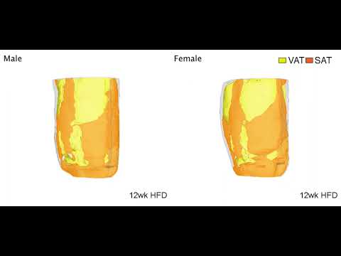
Watch a video of where males and female mice gain weight on a high-fat diet. While at some point females can have the same amount of visceral fat as males, there is still less inflammation. Credit: Alexis Stranahan, Medical College of Georgia
Stranahan acknowledges that the findings are potentially heretical and revolutionary and certainly surprising even to her. “We did these experiments to try and nail down, first of all, what happens first, the hormone perturbation, the inflammation, or the brain changes.”
To learn more about how the brain becomes inflamed, the researchers scrutinized increases in the amount and location of fat tissue as well as levels of sex hormones and brain inflammation in male and female mice at different time intervals as they grew fatter on a high-fat diet.
Since, much like with people, obese female mice tend to have more subcutaneous fat and less visceral fat than male mice, they reasoned that the distinctive fat patterns might be a key reason for the protection from inflammation the females enjoy before menopause.
In response to a high-fat diet, the investigators again found the distinctive patterns of fat distribution in males and females. They found no indicators of brain inflammation or insulin resistance, which also increase inflammation and can lead to diabetes, until after the female mice reached menopause. At about 48 weeks, menstruation stops and fat positioning on the females starts to shift somewhat, to become more like males.
They then compared the impact of the high-fat diet, which is known to increase inflammation body-wide, in mice of both sexes following surgery, similar to liposuction, to remove subcutaneous fat. They did nothing to directly interfere with normal estrogen levels, like removing the ovaries.
The subcutaneous fat loss increased brain inflammation in females without changing the levels of their estrogen and other sex hormones.
Bottom line: The females’ brain inflammation looked much more like the males’, including increased levels of classic inflammation promoters like the signaling proteins IL-1β and TNF alpha in the brain, Stranahan and her colleagues report.


Dr. Alexis Stranahan. Credit: Michael Holahan, Augusta University
“When we took subcutaneous fat out of the equation, all of a sudden the females’ brains start to exhibit inflammation the way that male brains do, and the females gained more visceral fat,” Stranahan says. “It kind of shunted everything toward that other storage location.” The transition occurred over about three months, which translates to several years in human time.
By comparison, it was only after menopause, that the females who did not have subcutaneous fat removed but did eat a high-fat diet, showed brain inflammation levels similar to the males, Stranahan says.
When subcutaneous fat was removed from mice on a low-fat diet at an early age, they developed a little more visceral fat and a little more inflammation in the fat. But Stranahan and her colleagues saw no evidence of inflammation in the brain.
One take-home lesson from the work: Don’t get liposuction and then eat a high-fat diet, Stranahan says. Another is: BMI, which simply divides weight by height and is commonly used to indicate overweight, obesity, and consequently increased risk of a myriad of diseases, is likely not a very meaningful tool, she says. An also easy and more accurate indicator of both metabolic risk and potentially brain health, is the also easy-to-calculate waist-to-hip ratio, she adds.
“We can’t just say obesity. We have to start talking about where the fat is. That is the critical element here,” Stranahan says.
She notes that the new study looked specifically in the hippocampus and hypothalamus of the brain. The hypothalamus controls metabolism and exhibits changes with inflammation from obesity that help control conditions that develop bodywide as a result. The hippocampus, a center of learning and memory, is regulated by signals associated with those pathologies but doesn’t control them, Stranahan notes. While these are good places to start such explorations, other regions of the brain could respond very differently, so she is already looking at the impact of loss of subcutaneous fat in others. Also, since her evidence indicates estrogen may not explain the protection females have, Stranahan wants to better define what does. One of her suspects is the clear chromosomal differences between the XX female and the XY male.
Stranahan has been studying the impact of obesity on the brain for several years and is among the first scientists to show that visceral fat promotes brain inflammation in obese male mice, and, conversely, transplanting subcutaneous fat reduces their brain inflammation. Females also have naturally higher levels of proteins that can tamp down inflammation. It’s been shown that in males, but not females, microglia, immune cells in the brain, are activated by a high-fat diet.
She notes that some consider the reason that females have higher stores of subcutaneous fat is to enable sufficient energy stores for reproduction, and she is not challenging the relationship. But many questions remain like how much fat is needed to maintain fertility versus the level that will affect your metabolism, Stranahan says.
Reference: ” Sex Differences in Adipose Tissue Distribution Determine Susceptibility to Neuroinflammation in Mice With Dietary Obesity” by Alexis M. Stranahan, De-Huang Guo, Masaki Yamamoto, Caterina M. Hernandez, Hesam Khodadadi, Babak Baban, Wenbo Zhi, Yun Lei, Xinyun Lu, Kehong Ding and Carlos M. Isales, 11 November 2022, Diabetes.
DOI: 10.2337/db22-0192
The research was supported by the National Institutes of Health (NIH).
Subcutaneous Fat Emerges as a Protector of Females’ Brains
Summary: Subcutaneous fat has a neuroprotective effect against brain inflammation, but the effect may diminish following menopause.
Source: Medical College of Georgia at Augusta University
Females’ propensity to deposit more fat in places like their hips, buttocks and the backs of their arms, so-called subcutaneous fat, is protective against brain inflammation, which can result in problems like dementia and stroke, at least until menopause, scientists report.
Males of essentially any age have a greater propensity to deposit fat around the major organs in their abdominal cavity, called visceral adiposity, which is known to be far more inflammatory. And, before females reach menopause, males are considered at much higher risk for inflammation-related problems from heart attack to stroke.
“When people think about protection in women, their first thought is estrogen,” says Alexis M. Stranahan, PhD, neuroscientist in the Department of Neuroscience and Regenerative Medicine at the Medical College of Georgia at Augusta University.
“But we need to get beyond the kind of simplistic idea that every sex difference involves hormone differences and hormone exposure. We need to really think more deeply about the underlying mechanisms for sex differences so that we can treat them and acknowledge the role that sex plays in different clinical outcomes.”
Diet and genetics are other likely factors that explain the differences broadly assigned to estrogen, says Stranahan, corresponding author of a study in the American Diabetes Association journal Diabetes.
She acknowledges that the findings are potentially heretical and revolutionary and certainly surprising even to her. “We did these experiments to try and nail down, first of all, what happens first, the hormone perturbation, the inflammation or the brain changes.”
To learn more about how the brain becomes inflamed, they looked at increases in the amount and location of fat tissue as well as levels of sex hormones and brain inflammation in male and female mice at different time intervals as they grew fatter on a high-fat diet.
Since, much like with people, obese female mice tend to have more subcutaneous fat and less visceral fat than male mice, they reasoned that the distinctive fat patterns might be a key reason for the protection from inflammation the females enjoy before menopause.
They found again the distinctive patterns of fat distribution in males and females in response to a high-fat diet. They found no indicators of brain inflammation or insulin resistance, which also increase inflammation and can lead to diabetes, until after the female mice reached menopause.
At about 48 weeks, menstruation stops and fat positioning on the females starts to shift somewhat, to become more like males.
They then compared the impact of the high-fat diet, which is known to increase inflammation body wide, in mice of both sexes following surgery, similar to liposuction, to remove subcutaneous fat. They did nothing to directly interfere with normal estrogen levels, like removing the ovaries.
The subcutaneous fat loss increased brain inflammation in females without moving the dial on levels of their estrogen and other sex hormones.
Bottom line: The females’ brain inflammation looked much more like the males’, including increased levels of classic inflammation promoters like the signaling proteins IL-1β and TNF alpha in the brain, Stranahan and her colleagues report.
“When we took subcutaneous fat out of the equation, all of a sudden the females’ brains start to exhibit inflammation the way that male brains do, and the females gained more visceral fat,” Stranahan says.
“It kind of shunted everything toward that other storage location.” The transition occurred over about three months, which translates to several years in human time.
By comparison, it was only after menopause, that the females who did not have subcutaneous fat removed but did eat a high-fat diet, showed brain inflammation levels similar to the males, Stranahan says.
When subcutaneous fat was removed from mice on a low-fat diet at an early age, they developed a little more visceral fat and a little more inflammation in the fat. But Stranahan and her colleagues saw no evidence of inflammation in the brain.
One take-home lesson from the work: Don’t get liposuction and then eat a high-fat diet, Stranahan says. Another is: BMI, which simply divides weight by height and is commonly used to indicate overweight, obesity and consequently increased risk of a myriad of diseases, is likely not a very meaningful tool, she says.
An also easy and more accurate indicator of both metabolic risk and potentially brain health, is the also easy-to-calculate waist to hip ratio, she adds.
“We can’t just say obesity. We have to start talking about where the fat is. That is the critical element here,” Stranahan says.
She notes that the new study looked specifically in the hippocampus and hypothalamus of the brain. The hypothalamus controls metabolism and exhibits changes with inflammation from obesity that help control conditions that develop bodywide as a result.
The hippocampus, a center of learning and memory, is regulated by signals associated with those pathologies but doesn’t control them, Stranahan notes. While these are good places to start such explorations, other regions of the brain could respond very differently, so she is already looking at the impact of loss of subcutaneous fat in others.
Also, since her evidence indicates estrogen may not explain the protection females have, Stranahan wants to better define what does. One of her suspects is the clear chromosomal differences between the XX female and the XY male.
Stranahan has been studying the impact of obesity on the brain for several years and is among the first scientists to show that visceral fat promotes brain inflammation in obese male mice, and, conversely, transplanting subcutaneous fat reduces their brain inflammation.
See also


Females also have naturally higher levels of proteins that can tamp down inflammation. It’s been shown that in males, but not females, microglia, immune cells in the brain, are activated by a high-fat diet.
She notes that some consider the reason that females have higher stores of subcutaneous fat is to enable sufficient energy stores for reproduction, and she is not challenging the relationship. But many questions remain like how much fat is needed to maintain fertility versus the level that will affect your metabolism, Stranahan says.
Funding: The research was supported by the National Institutes of Health.
About this neuroscience research news
Author: Toni Baker
Source: Medical College of Georgia at Augusta University
Contact: Toni Baker – Medical College of Georgia at Augusta University
Image: The image is in the public domain
Original Research: Closed access.
“Sex Differences in Adipose Tissue Distribution Determine Susceptibility to Neuroinflammation in Mice With Dietary Obesity” by Alexis M. Stranahan et al. Diabetes
Abstract
Sex Differences in Adipose Tissue Distribution Determine Susceptibility to Neuroinflammation in Mice With Dietary Obesity
Preferential energy storage in subcutaneous adipose tissue (SAT) confers protection against obesity-induced pathophysiology in females. Females also exhibit distinct immunological responses, relative to males. These differences are often attributed to sex hormones, but reciprocal interactions between metabolism, immunity, and gonadal steroids remain poorly understood.
Here, we systematically characterized adipose tissue hypertrophy, sex steroids, and inflammation in male and female mice after increasing durations of high-fat diet (HFD)-induced obesity.
After observing that sex differences in adipose tissue distribution before HFD were correlated with lasting protection against inflammation in females, we hypothesized that a priori differences in the ratio of subcutaneous to visceral fat might mediate this relationship.
To test this, male and female mice received SAT lipectomy (LPX) or sham surgery before HFD challenge, followed by analysis of glial reactivity, adipose tissue inflammation, and reproductive steroids.
Because LPX eliminated female resistance to the pro-inflammatory effects of HFD without changing circulating sex hormones, we conclude that sexually dimorphic organization of subcutaneous and visceral fat determines susceptibility to inflammation in obesity.
Females Found to Itch Less Than Males
Summary: The female hormone estradiol helps suppress itch associated with psoriasis. The findings shed light on why men are more prone to psoriasis and offers hope for new targeted treatment for itch disorders.
Source: Kyoto University
Among the many reasons men may have for envying women, at least when it comes to skin inflammation, is that women have a significantly lower incidence of severe psoriasis. However, the underlying reason for the sex differences had been unclear.
Now, a team of researchers has found that the female hormone estradiol suppresses psoriasis, and the protective role of the hormone has provided a basis for its therapeutic potential.
“Our results have not only revealed the molecular mechanisms of sex differences in psoriasis but also shed new light on our understanding of the physiological role of estradiol,” says Hamamatsu University School of Medicine’s Tetsuya Honda, formerly of Kyoto University.
The team tested conditional knockout mice, or cko mice, with ovaries removed but supplemented with estradiol pellets or a placebo. In contrast to wild-type mice, the cko mice without the natural ovarian hormones estradiol showed symptoms of severe skin inflammation.
Once these mice were given estradiol, the production of IL-17A and IL-1β cytokines in neutrophil and macrophage immune cells was reversed, reducing the inflammation. This effect was also observed in human neutrophils in vitro.
What intrigued the researchers was how the lack of estrogen receptors in immune cells made estradiol ineffective against the cytokines.
“These results indicate that estradiol suppresses psoriatic inflammation by regulating neutrophil and macrophage cells,” concludes the author.
About this neuroscience research news
Author: Press Office
Source: Kyoto University
Contact: Press Office – Kyoto University
Image: The image is credited to Kyoto University
Original Research: Closed access.
“Estradiol suppresses psoriatic inflammation in mice by regulating neutrophil and macrophage functions” by Akimasa Adachi et al. Journal of Allergy and Clinical Immunology
Abstract
See also

Estradiol suppresses psoriatic inflammation in mice by regulating neutrophil and macrophage functions
Background
Psoriasis is a common inflammatory skin disease resulting from dysregulation of the IL-23/TH17 immune axis. The prevalence and severity of psoriasis is higher in men than in women, although the underlying reasons for this are unclear.
Objective
We studied whether estradiol, a female hormone, plays protective roles in imiquimod-induced psoriatic inflammation in mice by regulating neutrophil and macrophage functions.
Methods
Wild-type mice and conditional knockout mice were ovariectomized, supplemented with placebo or estradiol pellets, and an imiquimod-containing cream applied.
Results
Mice without endogenous ovarian hormones exhibited exacerbated psoriatic inflammation including increased production of IL-17A and IL-1β, which was reversed by exogenously added estradiol. The suppressive effect of estradiol on the production of IL-1β and IL-17A was abolished in mice lacking estrogen receptors in neutrophils and macrophages (Esr1f/fEsr2f/fLysM-Cre+ mice). IL-1β, which is required for production of IL-17A in the psoriasis model, was mainly produced by neutrophils and inflammatory macrophages. Estradiol suppressed IL-1β production from neutrophils and macrophages in mice both in vivo and in vitro and from human neutrophils in vitro.
Conclusion
Our results suggest a novel mechanism for sex-dependent differences in psoriasis clinical phenotypes that may shed new light on the pathology of psoriasis.
See Male Spiders Catapult to Escape Being Devoured by Females After Sex
Two Philoponella prominens spiders mating. If the male is fast enough, it will catapult itself to safety.
Shichang Zhang
Some female spiders have a reputation of eating their mates after copulation. But some male orb weaver spiders have worked out a dramatic survival mechanism: catapulting themselves to safety at high speeds.
A team of researchers led by ecologist Shichang Zhang of Hubei University in China published a study on the spiders’ energetic escapes in the journal Current Biology on Monday.
Male Philoponella prominens orb weaver spiders are the kangaroos of the arachnid world.
“Using a mechanism that hadn’t been described before, the male spiders use a joint in their first pair of legs to immediately undertake a split-second catapult action, flinging themselves away from their partners at impressive speeds clocked at up to 88 centimeters per second (cm/s),” Cell Press, the publisher of Current Biology, said in a news release Monday.
The researchers captured video of the catapulting action that shows the males’ quick getaway method.
Males that didn’t immediately catapult away after sex were caught and eaten “in an act of sexual cannibalism.” The discovery came about as the team studied sexual selection in the orb weavers, which live in large communities together. They observed 155 successful matings with 152 ending in a catapult to freedom. The three that didn’t catapult became dinner.
To test the observations, the researchers prevented 30 males from catapulting away. Those males also became dinner.
“Females may use this behavior to judge the quality of a male during mating,” Zhang said. “If a male could not perform catapulting, then kill it, and if a male could perform it multiple times, then accept its sperm.”
It may be a spider-eat-spider world out there, but at least some of the arachnids have figured out the secret to survival. It requires strong legs and good timing, which is life advice that could apply to a lot of us.
These Male Spiders Evolved a Post-Sex Catapult to Escape Cannibalistic Females
Researchers in China have described yet another freaky sex habit of spiders (as if there weren’t enough already): Some male spiders launch themselves at great speed off their cannibalistic female partners, to avoid being eaten after copulating. The way they make their egress is similar to the mechanism at play in catapults, according to the new study.
The spiders are communal orb-weaving spiders (Philoponella prominens), and they have active and potentially fatal sex lives. Like praying mantises, the female arachnids have an appetite for their sex partners. But the 0.12-inch (3 mm) males of the species have developed an escape plan: They capitalize on an adaptation in the joint in their front two legs to launch themselves off the females, at speeds of nearly 3 feet (88.2 cm) per second. The team’s research is published today in Current Biology.
“Males can use super-fast actions with extraordinary kinetic performance to escape the female’s attack,” said Shichang Zhang, a behavioral ecologist at Hubei University, in an email to Gizmodo. “This may help scientists to consider the balance or trade-off between cost in physical strength and the benefit of paternity when studying sexual conflict.”
There are many other methods male spiders use to counter sexual cannibalism, Zhang said, including nuptial gifts, pretending to be dead, and cutting off their own genitals or mutilating the females’; they’re as cutthroat as they are creative. But the catapulting approach is new to the researchers.
In a lab, the team mated 155 pairs of spiders; in 152 of the encounters, the males catapulted off the females to safety. The three males that did not do the behavior were captured, killed, and eaten by the females.
The researchers attributed the males’ ability to launch off their sexual partners to a leg joint called the tibia-metatarsus. The tibia-metatarsus (and all the leg joints in the spiders) are ensconced in sheathes called thecae, which increase the limbs’ elasticity. In the front two legs of the male spiders, the surface area of the thecae was much larger than on the other legs.
These spiders don’t have sex like quite humans: Mating lasts about 30 seconds, and male spiders use an appendage called the palp to inject sperm into the female’s epigynum, a hard plate on the bottom of the abdomen.
The females’ eggs aren’t immediately fertilized. Instead, the female can store sperm and only release the egg for fertilization when it is ready. It can also squeeze the sperm out or kill them if the sperm (and the male who provided them) are found wanting.
“Mating is ended by the female; once [they] sense the aggressiveness of the female, males catapult off, but if a male can not sense the danger, it may not catapult before the female kills it,” Zhang said.
“Through the catapulting, male can escape female sexual cannibalism, and female can choose males with high quality, because the kinetic performance may directly correlate with male’s physical condition. Only those with good quality can catapult off far or can catapult for several times,” Zhang added.
The team also observed that the males employed a silk ‘safety line’ to dangle near the webs where the mating occurred, which they believe was a means for the male to return in case they wanted to attempt mating again. On a human scale, according to Zhang, the action is equivalent to a 5-foot-10 human leaping a third of a mile from their partner after sex. You know, to be safe.
More: Researchers Found Glowing Spider Fossils in the South of France
Researchers Discover Dolphin Females Have Working Clitoris – “Surprisingly Similar” to the Shape in Humans
Dolphins in action. Credit: Dara Orbach
Like humans, female dolphins have a functional clitoris, according to a study appearing today (January 10, 2022) in the journal Current Biology. The findings are based on the discovery that the clitoris-like structure positioned in the vaginal entrance of bottlenose dolphins has lots of sensory nerves and erectile bodies.
“The dolphin clitoris has many features to suggest that it functions to provide pleasure to females,” says first author Patricia Brennan, an assistant professor of biological sciences at Mount Holyoke College in Massachusetts.
Scientists have known that dolphins are highly social. They have sex throughout the year as a way of forging and maintaining social bonds. It had been noted also that dolphin females have a clitoris in the vagina in a spot that would make stimulation during copulation likely. There’ve also been reports of females rubbing each other’s clitorises with their snouts, flippers, and flukes.


This photograph depicts dolphin arousal. Credit: Dara Orbach
In the new study, Brennan and colleagues decided to take a closer look at the dolphin clitoris. They looked carefully at clitorises from 11 females that had died naturally. They examined them for the presence, shape, and configuration of erectile bodies. They also looked at how nerve fibers ran through the tissues. What they saw supports the notion of a working clitoris in dolphins.
“Just like the human clitoris, the dolphin clitoris has large areas of erectile tissue that fill up with blood,” Brennan says.
The erectile tissue shape changes as animals become adults, she added, suggesting that it acquires a functional role. The studies further show that the clitoris body has large nerves and many free nerve endings right underneath the skin, which is much thinner there than in the adjacent skin. They also found genital corpuscles much like those previously described in the human clitoris and penis tip, which are known to be involved in the pleasure response.

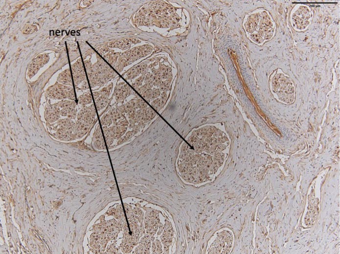
This image depicts the big nerves in the dolphin clitoris. Credit: Patricia Brennan
Overall, Brennan says that the erectile bodies in dolphins are “surprisingly similar” to the shape of the erectile bodies in humans.
“Since the entire pelvis of dolphins is so different to humans, it was surprising to see how similar the shapes were,” she says. “Also, the size of the nerves in the clitoris body was very surprising. Some were larger than half a millimeter in diameter.”
Brennan said they got curious about the dolphin clitoris while studying the evolution of vaginas in dolphins.
“Every time we dissected a vagina, we would see this very large clitoris, and we were curious whether anyone had examined it in detail to see if it worked like a human clitoris,” she says. “We knew that dolphins have sex not just to reproduce, but also to solidify social bonds, so it seemed likely that the clitoris could be functional.”
The researchers note that there’s been little study of the clitoris and female sexual pleasure in nature. In fact, even the human clitoris wasn’t fully described until the 1990s.
“This neglect in the study of female sexuality has left us with an incomplete picture of the true nature of sexual behaviors,” Brennan says. “Studying and understanding sexual behaviors in nature is a fundamental part of understanding the animal experience and may even have important medical applications in the future.”
Her team will continue to examine the clitoris and genitalia of dolphins and many other vertebrates to help fill in these gaps.
Reference: “Evidence of a functional clitoris in dolphins” by Patricia L.R. Brennan, Jonathan R. Cowart and Dara N. Orbach, 10 January 2022, Current Biology.
DOI: 10.1016/j.cub.2021.11.020
The Dolphin Clitoris Is Full of Surprises, Scientists Discover
The bottlenose dolphin (Tersiops truncatus) appears to have a very large and well-developed clitoris, potentially better placed for coital pleasure than the clitoris of humans, according to new research.
The visible tip of the human clitoris is but the size of a pea and located slightly north of the vagina and urethra (although much of the structure remains hidden in the pelvis or under a ‘hood’ of skin).
The head of the dolphin clitoris, on the other hand, is slightly larger and located right near the vagina entrance. What’s more, the whole organ has an ‘S’-shaped bend in it, which suggests it can stick out even further when erect.
During copulation, it would be almost impossible for a dolphin penis to avoid, experts say.
“The dolphin clitoris has many features to suggest that it functions to provide pleasure to females,” says biologist Patricia Brennan from Mount Holyoke College in Massachusetts.
“We knew that dolphins have sex not just to reproduce, but also to solidify social bonds, so it seemed likely that the clitoris could be functional.”
Computer reconstruction of the dolphin clitoris. (Dara Orbach/Mount Holyoke College)
Today, we still know very little about the human clitoris and even less about its counterpart in other species. All female mammals are known to have a clitoris-like structure, but we still aren’t sure how these organs function or if they give animals pleasure.
Like humans, female dolphins are known to copulate all year round, but only sometimes are they ovulating. This suggests the species mates for more than just reproduction.
In the wild, for instance, bottlenose dolphins have been observed partaking in group orgies, where male and females alike use their snouts, flippers, and flukes to rub the protruding clitorises and penises of their peers.
Direct stimulation of the clitoris has also been observed in sexual interactions between only females.
Unfortunately, we can’t scan a dolphin’s brain during all this hanky panky to see if these creatures really are having fun, so researchers have turned to the clitoris itself for answers.
When scanning the sexual organs of 11 naturally deceased female dolphins, the team found an abundance of erectile tissue, blood vessels, and nerve endings in the clitoris.
Similar to the human clitoris, the glans of the dolphin clitoris is also enclosed in a hood. In dolphin adulthood, this hood becomes wrinkled, possibly allowing the tip of the organ, which includes erectile tissue, to swell with blood when aroused.
Arteries in the clitoris were also found to closely trace clitoral nerves, which is an indication of orgasm function in humans.
“Since the entire pelvis of dolphins is so different to humans, it was surprising to see how similar the shapes were,” says Brennan.
“Also, the size of the nerves in the clitoris body was very surprising. Some were larger than half a millimeter in diameter.”
Given that the penis and the clitoris develop from the same structures, the findings could help explain why dolphins of all sexes have been seen masturbating on the sandy floor. Some have even been caught using ‘sex toys’, in the form of dead fish or wriggling eels.
Dolphin sex is clearly a kinky affair, and it’s drawn the interest of researchers for years now. Still, most experts have been interested in the dolphin penis, investigating what it looks like, and examining how it fits with the dolphin vagina.
In comparison, the dolphin clitoris has been all but overlooked. And that’s the case for most female mammals.
“Very little is known about female reproductive morphology in most wild vertebrate species,” said researcher Dara Orbach, when announcing the preliminary results of her dolphin dissection in 2019.
“This research provides a comparative framework to explore other functions of sex that may not be unique to humans.”
If sexual pleasure really does hold evolutionary significance, female pleasure among mammals might tell us how. Ignoring this side of sex will give us only half the picture, and as we all know, it takes at least two to tango.
The study was published in Current Biology.