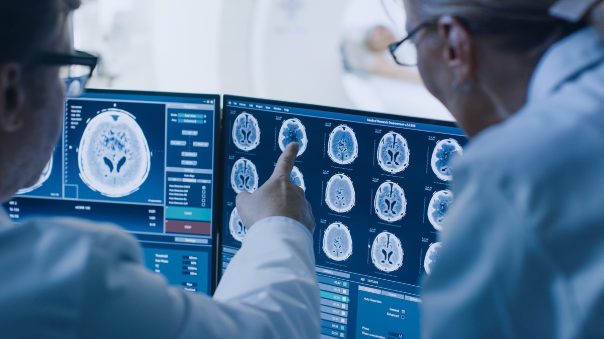
- Scientists ‘switch off’ autism symptoms using $3 epilepsy drug: discovery New York Post
- Scientists ‘CURE autism’ using $3 epilepsy drug in mice – in potential breakthrough Daily Mail
- Drug Shown to Alleviate Autism-Associated Behavior Neuroscience News
- MYT1L haploinsufficiency in human neurons and mice causes autism-associated phenotypes that can be reversed by genetic and pharmacologic intervention | Molecular Psychiatry Nature.com
- Autism-linked MYT1L mutations prompt ‘identity crisis’ in budding brain cells | Spectrum Spectrum – Autism Research News
- View Full Coverage on Google News
Tag Archives: Epilepsy
Fear of Public Places Is Common in Adults With Epilepsy
Summary: Phobic and agoraphobic symptoms are common in those with epilepsy and result in a poorer quality of life.
Source: Wake Forest University
About 5.1 million people in the U.S. have a history of epilepsy, which causes repeated seizures. According to the Epilepsy Foundation, epilepsy is the fourth most common neurological disorder.
While current research has shown an increase in anxiety and depression among people with epilepsy, little is known about this population and agoraphobia, an anxiety disorder that involves the fear of being in a public place or in a situation that might cause panic or embarrassment.
However, a recent study from Heidi Munger Clary, M.D., M.P.H., associate professor of neurology at Wake Forest University School of Medicine, shows that phobic and agoraphobic symptoms are common and associated with poor quality of life in people with epilepsy.
The study appears online in Epilepsy Research.
“We know that agoraphobia can lead to delays in patient care because of a reluctance to go out in public, which includes appointments with health care providers,” said Munger Clary, the study’s principal investigator. “So, this is an area that needs more attention in clinical practice.”
In the study, researchers conducted a cross-sectional analysis of baseline clinical data from a neuropsychology registry cohort study. Researchers analyzed a diverse sample of 420 adults, ages 18 to 75, with epilepsy who underwent neuropsychological evaluation over a 14-year period at Columbia University Medical Center in New York.
“More than one-third of the participants reported significant phobic/agoraphobic symptoms,” Munger Clary said. “We also found that phobic/agoraphobic symptoms, along with depression symptoms, were independently associated with poor quality of life, but generalized anxiety symptoms were not.”
According to Munger Clary, because phobic/agoraphobic symptoms are not routinely assessed by clinicians, the findings may suggest a need for future studies to develop more comprehensive screeners for psychiatric comorbidity in epilepsy.
“Symptoms of agoraphobia do not fully overlap with generalized anxiety or depression symptoms that are often screened in routine practice,” Munger Clary said.
“Providers might want to consider more robust symptom screening methods to identify and better assist these patients. This may be important to improve health equity, given other key study findings that show those with lower education and non-white race/ethnicity had increased odds of significant phobic/agoraphobic symptoms.”
Funding: This work was supported in part by the National Institutes of Health under grants R01 NS035140, KM1 CA156709, UL1 TR001420 and 5KL2TR001421-04.
About this epilepsy and psychology research news
Author: Myra Wright
Source: Wake Forest University
Contact: Myra Wright – Wake Forest University
Image: The image is in the public domain
See also


Original Research: Open access.
“Afraid to go out: Poor quality of life with phobic anxiety in a large cross-sectional adult epilepsy center sample” by Munger Clary et al. Epilepsy Research
Abstract
Afraid to go out: Poor quality of life with phobic anxiety in a large cross-sectional adult epilepsy center sample
Purpose
People with epilepsy (PWE) have unmet healthcare needs, especially in the context of mental health. Although the current literature has established increased incidence of anxiety and depression in PWE and their contribution to poor quality of life, little is known regarding the presence and impact of specific phobia and agoraphobia. Our aim was to assess factors associated with high phobic/agoraphobic symptoms in a large, single tertiary epilepsy center sample, and to assess their impact on quality of life.
Methods
In a diverse sample of 420 adults with epilepsy, cross-sectional association of demographic, epilepsy and cognitive factors with high phobic symptoms were assessed using multiple logistic regression. Symptoms were measured with the SCL-90R validated self-report subscale (T-score ≥ 60 considered high phobic symptom group). Multiple logistic regression modeling was used to assess for independent association of demographic and clinical variables with presence of high phobic symptoms, and multiple linear regression modeling was used to evaluate for independent cross-sectional associations with epilepsy-specific quality of life (QOLIE-89).
Results
Lower education (adjusted OR 3.38), non-White race/ethnicity (adjusted OR 2.34), and generalized anxiety symptoms (adjusted OR 1.91) were independently associated with high phobic/agoraphobic symptoms, all p < 0.005. Phobic/agoraphobic symptoms were independently associated with poor quality of life as were depression symptoms, older age, and non-White race/ethnicity. Generalized anxiety did not demonstrate a significant independent association with quality of life in the multivariable model.
Conclusion
In this study sample, phobic/agoraphobic symptoms were independently associated with poor quality of life. Clinicians should consider using more global symptom screening instruments with particular attention to susceptible populations, as these impactful symptoms may be overlooked using generalized-anxiety focused screening paradigms.
A Person Who Lived 800 Years Ago Is the Origin of a Modern Seizure Disorder, Scientists Say
Scientists in Australia believe that they’ve discovered the centuries-old origins of a rare form of childhood epilepsy caused by a genetic mutation: a single common ancestor who lived in Britain roughly 800 years ago. The find is especially notable because hereditary conditions of this kind typically don’t survive for so long in the population.
Epilepsy is a broad term for recurring bursts of abnormal brain activity that trigger neurological symptoms, most prominently seizures. It can have many different causes, including variations in our genes passed down between families. When these seizures are accompanied by fever, they’re also known as febrile seizures.
This new study, led by researchers at the University of Melbourne’s Epilepsy Research Center, looked at cases of childhood febrile seizures strongly tied to the SCN1Bc.363C>G variant. This variant has been found among multiple unrelated families in Australia, the UK, and the U.S. Many of the families had a long history of early epilepsy, and the disorder appears to be a dominant genetic condition, meaning a disease that can be caused by only having one copy of the bad gene. But the researchers were curious whether this mutation had been passed down by a lone common ancestor to these affected families or if it had independently arisen multiple times in human history.
The group tried to trace back the lineage of the SCN1Bc.363C>G variant in 14 families with these seizures. They also analyzed genome data from the UK Biobank, a large-scale and long running study of people’s health that also collects their genetic information.
Within the biobank, the researchers identified another 74 individuals with the same variant. And all of these people had similar patterns of other genetic variations surrounding the variant—a grouping of genes that’s known as a haplotype. It’s very unlikely that all of these people would have the same common haplotype without having some shared ancestry, the researchers say, meaning that the existence of this genetic disease today is probably due to just one ancestor, also known as a founder event. And as near as they can tell, this ancestor lived about 800 years ago.
“Here, we report evidence of a single founder event giving rise to the SCN1Bc.363C>GQ11variant in 14 independent families with epilepsy,” the authors wrote in their paper, published Tuesday in The American Journal of Human Genetics.
There are other genetic disorders or traits that can be cleanly traced back to a single founder event. But these disorders tend to appear later in life (after a person has already reproduced) or to be recessive, meaning that they only cause disease when someone inherits both copies of the bad variant. So it’s very unusual to see the same with a damaging dominant mutation that shows up in childhood. Often, these mutations are weeded out in a short time, since affected people would be less likely to survive into adulthood and pass on the mutation to the next generation—an example of natural selection.
This mutation, the authors speculate, might have endured because most people with it experience relatively mild seizures. Only about 70% of people with the mutation seem to become sick at all, something known as incomplete penetrance. In other words, this mutation might cause trouble, but not enough to have kept people who had it from living their lives and passing on their genes.
Aside from learning more about this disease, the authors say their findings could have broader implications. There may very well be other genetic mutations out there that similarly linger in the population at low levels but which might actually turn out to be more harmful than currently assumed.
“These findings suggest variants present in the population at low frequencies should be considered potentially pathogenic in mild phenotypes with incomplete penetrance and may be more important than previously thought,” they wrote.
Peptide Delivered by Nasal Spray Can Reduce Seizure Activity and Protect Neurons in Alzheimer’s and Epilepsy
Summary: A1R-CT, a novel peptide that binds to neurabin, can be administered via a nasal spray and holds the potential to interrupt uncontrollable brain activity associated with TBI, stroke, epilepsy, and Alzheimer’s disease.
Source: Medical College of Georgia at Augusta University
A novel peptide augments the brain’s natural mechanism to help prevent seizures and protect neurons in research models of both Alzheimer’s and epilepsy, scientists report.
The A1R-CT peptide the scientists developed, which can be administered through a nasal spray, holds promise for tamping down the uncontrolled electrical activity that is common after traumatic brain injury, stroke and which affects more than half of individuals with Alzheimer’s, says Dr. Qin Wang, neuropharmacologist and founding director of the Program for Alzheimer’s Therapeutics Discovery at the Medical College of Georgia at Augusta University.
The fact that it can be delivered through the nose indicates the peptide’s potential as a new seizure rescue medication as well, to help interrupt, for example, a seizure cluster, where disabling seizures are occurring back-to- back, says Wang, corresponding author of the study in the journal JCI Insight.
A1R-CT works by inhibiting neurabin, a protein that helps ensure that the protective mechanism itself, which tamps down the hyperexcitability of neurons that disrupts normal communication and produces seizures, doesn’t overdo, she says.
The peptide was named after the protective adenosine 1 receptor on the surface of neurons, which gets activated by adenosine, a chemical made mostly in the brain by neuron-supporting glial cells in response to hyperexcitability.
“This is a powerful receptor to then silence the neurons,” Wang says. This natural, calming relationship also is known to block electrical activity that can result in an irregular heartbeat. In fact, an injectable form of adenosine is used to treat a very high heart rate.
“But the A1 receptor itself has to be regulated because if it’s activated too much, you will fall asleep,” says Wang. “The neurons try to make sure everything stays in control and in most of us, it works pretty well. We don’t fall asleep at our desk. We don’t have seizures,” she says, noting that caffeine blocks the A1 receptor.
Alzheimer’s often is accompanied by seizures because the characteristic buildup of the proteins amyloid and tau in the brain disrupts communication between neurons, creates increased oxidative stress and resulting inflammation, and in response to the altered dynamic, neurons can become hyperexcited, she says.
“In Alzheimer’s there are so many things that go wrong,” she says. Seizures can precede the cognitive decline in Alzheimer’s, and definitely contribute to it, Wang says.
A1 receptor’s activation by adenosine in this type of hyperactive scenario makes it seem like a logical treatment target for seizures. But the fact that it is so pervasive throughout the body, including in the heart, lungs and kidneys, makes potential extensive side effects likely.
Back to neurons’ desire for homeostasis, Wang and her colleagues were the ones who found that the protein neurabin, which appears to be primarily present in the brain, provides that balance to prevent hyperactivity of the A1 receptor.
The fact neurabin is primarily in the brain, means that altering its activity should not have the potential body-wide impact of directly altering A1 receptor activity, Wang says.
“Neurabin is a brake so it doesn’t do too much,” Wang says. “But now we need to remove it to unleash A1’s power.”
So, they set to work developing the peptide that could instead interfere with the A1 receptor and neurabin’s interaction and so enable more of the natural protective, seizure-reducing benefit.
A1 receptor activation tamps down the excited state of the neurons by modulating ion channels — proteins in the cell membrane that allow passage through the cell of other proteins — which help generate electrical signals.
A result is so-called hyperpolarization, which means the neuron is less likely to fire an electrical signal.
“The more polarized the neurons are, the harder it is for them to get excited,” Wang says.
A1 receptor activation also decreases the release of glutamate, a neurotransmitter produced by neurons that excites neurons. It also provides additional benefit to neurons by providing some protection from inadequate oxygen and blood supplies, which may occur in the case of an injury. The scientists have noted a dramatic reduction in death of neurons in their Alzheimer’s model, for example, with the use of their peptide.
Now they’ve shown that inhibiting neurabin — either by reducing it directly or with their peptide — enables increased action by A1C to reduce excessive electrical activity in the brain. They’ve shown the peptide is effective in both a mouse model of severe seizures and seizures in an Alzheimer’s mouse model. And It’s effective when directly injected into the brain or via nasal spray.
The scientists opted to look at nasal spray delivery to fully explore the peptide’s potential clinical benefit. They found a similar robust response in both the seizure and Alzheimer’s models.
Looking further at the impact of targeting neurabin, they found that mice with a neurabin deficiency had significantly shorter, less severe seizures and they all survived. Those with the normal neurabin levels intact experienced seizures lasting for up to 30 minutes and about 10% of the mice died shortly afterward.
Blocking A1 receptor resulted in more severe seizures in the neurabin-deficient mice and increased the death rate to more than 50%.
Next steps include additional exploration of ideal doses and delivery times for specific conditions the peptide may be used to treat.
The scientific team also continues to tweak the peptide to ensure it functions optimally, and is pursuing funding needed to pursue clinical trials.
Wang, a Georgia Research Alliance Eminent Scholar, came to MCG in April 2021 from the University of Alabama at Birmingham, where she began the studies of A1 receptor and peptide development. She continues extensive collaboration with her colleagues at UAB on the studies who are coauthors on the new paper. First author Dr. Shalini Saggu is now also a faculty member in the MCG Department of Neuroscience and Regenerative Medicine.
Epileptic seizures are common after a traumatic brain injury; a stroke, which is considered an acquired brain injury; and with chronic neurodegenerative diseases including Alzheimer’s.
See also

As much as 64% of the some 50 million individuals with Alzheimer’s experience seizures, the scientists write. Patients can experience generalized tonic-clonic seizures, in which they fall down, shake and become unresponsive. Also, focal onset seizure, which tend to be shorter and may include repetitive movement of the arms or legs, lip smacking and chewing.
Seizures are uncontrolled in about 40% of people, which indicates an urgent need for novel therapies, the scientists write, and current therapies tend to be less effective in individuals with Alzheimer’s. Left uncontrolled, seizures can produce brain damage and cognitive impairment.
Adenosine also is a building block of our DNA and a component of the cell fuel ATP.
Funding: The research was supported by the National Institutes of Health.
About this neuropharmacology research news
Author: Toni Baker
Source: Medical College of Georgia at Augusta University
Contact: Toni Baker – Medical College of Georgia at Augusta University
Image: The image is in the public domain
Original Research: Open access.
“A peptide blocking the ADORA1-neurabin interaction is anticonvulsant and inhibits epilepsy in an Alzheimer’s model” by Qin Wang et al. JCI Insights
Abstract
A peptide blocking the ADORA1-neurabin interaction is anticonvulsant and inhibits epilepsy in an Alzheimer’s model
Epileptic seizures are common sequelae of stroke, acute brain injury, and chronic neurodegenerative diseases, including Alzheimer’s disease (AD), and cannot be effectively controlled in approximately 40% of patients, necessitating the development of novel therapeutic agents.
Activation of the A1 receptor (A1R) by endogenous adenosine is an intrinsic mechanism to self-terminate seizures and protect neurons from excitotoxicity. However, targeting A1R for neurological disorders has been hindered by side effects associated with its broad expression outside the nervous system.
Here we aim to target the neural-specific A1R/neurabin/regulator of G protein signaling 4 (A1R/neurabin/RGS4) complex that dictates A1R signaling strength and response outcome in the brain. We developed a peptide that blocks the A1R-neurabin interaction to enhance A1R activity. Intracerebroventricular or i.n. administration of this peptide shows marked protection against kainate-induced seizures and neuronal death.
Furthermore, in an AD mouse model with spontaneous seizures, nasal delivery of this blocking peptide reduces epileptic spike frequency. Significantly, the anticonvulsant and neuroprotective effects of this peptide are achieved through enhanced A1R function in response to endogenous adenosine in the brain, thus, avoiding side effects associated with A1R activation in peripheral tissues and organs.
Our study informs potentially new anti-seizure therapy applicable to epilepsy and other neurological illness with comorbid seizures.
Epilepsy drug ‘doubles children’s autism risk’ if their mother takes it while pregnant
Epilepsy drug ‘doubles children’s autism risk’ if their mother takes it while pregnant
A drug used to prevent epileptic seizures and migraine has been found to double the chances of a child developing autism if their mother takes it while pregnant.
An urgent review has been launched into topiramate, known by the brand name Topamax, which has been prescribed for decades.
The Medicines and Healthcare products Regulatory Agency (MHRA) has begun a safety review to assess the ‘benefits and risks’ of the drug, which is likely to raise the odds of other intellectual impairments, as well as congenital birth defects.


A drug used to prevent epileptic seizures and migraine has been found to double the chances of a child developing autism if their mother takes it while pregnant. A file photo is used above
It follows warnings about another epilepsy drug, sodium valproate, marketed as Epilim, which has been linked to higher-than-normal rates of the same conditions.
Seven years ago the MHRA ordered that women of child-bearing age should be warned about sodium valproate’s risks. However, pregnant women were still being prescribed it earlier this year. Experts believe 20,000 babies have been harmed as a result.
The MHRA launched its investigation late last month, after Scandinavian scientists conducted an observational study looking at rates of autism and intellectual disability in children whose mothers took topiramate while pregnant.
They found that about three per cent of such children had an autism spectrum disorder (ASD) diagnosis – double the 1.5 per cent rate among those not exposed to the drug.


An urgent review has been launched into topiramate, known by the brand name Topamax, which has been prescribed for decades. A file photo is used above
Some 3.5 per cent of children whose mothers took it while pregnant were diagnosed with an intellectual disability – about four times higher than the rate of 0.8 per cent among those unexposed. The results were derived from looking at patient records from 4.5 million children across five Nordic countries, of which almost 25,000 had been exposed to topiramate in the womb.
Writing in the Journal Of The American Medical Association Neurology, the scientists warned: ‘Our results do not suggest that topiramate is a safe alternative to sodium valproate (Epilim).’
Drugs firm Janssen, which makes topiramate / Topamax, said packs already contained a warning that it ‘should not be used during pregnancy unless the potential benefit outweighs the potential risk’.
Epilepsy and Hearing Loss Could Be the First Signs of Parkinson’s
Keeping an eye on developing certain health conditions is a part of the aging process everyone faces at some point, whether it’s heart disease, dementia, or diabetes. But with more than 1 million confirmed cases and 60,000 patients in the U.S. diagnosed a year, Parkinson’s disease is another potential concern that shouldn’t be overlooked. Fortunately, mounting research is providing the medical community with a better understanding of how doctors can treat the neurological condition and any red flags that could help them detect and diagnose it as soon as possible. And according to a new study, two particular symptoms could be the first signs of Parkinson’s disease. Read on to see the health signals that warrant a visit to your doctor.
RELATED: If You’ve Done This, Your Parkinson’s Risk Goes Up 90 Percent, Study Says.


In a new study published in JAMA Neurology, a team of researchers from Queen Mary University of London aimed to shed more light on which health problems might be precursors or early warning signs of Parkinson’s disease. To do this, the group analyzed the health records of 1,016,277 people who lived in East London between 1990 and February 2018, including 1,055 who developed Parkinson’s throughout the record-keeping period.
Results showed that typical warning signs such as tremors or memory loss could be seen in patients five to ten years before diagnosis. But researchers found two other early symptoms to be reliable warning signs of the condition: epilepsy and hearing loss.


According to the study, results found that hearing loss signaled a 66 percent increased risk of patients developing Parkinson’s disease, with researchers speculating that it could result from the beginning of changes in the brain’s functions. “Although the role of early hearing loss requires further research, it is possible that this factor represents another deficit in sensory processing that occurs as part of Parkinson’s disease pathogenesis, similar to visual impairment,” Cristina Simonet, MD, a neurologist, PhD student, and the study’s lead author, said in a statement.
But the association with epilepsy was even more significant, with patients who experienced the condition found to be 2.5 times more likely to develop Parkinson’s later in life. The researchers pointed out that previous studies have established a connection between epilepsy medication and Parkinson’s, providing possible causality that required further investigation. “The prevalence of epilepsy in patients with Parkinson’s disease has been reported as being higher than the estimated prevalence in the general population,” the researchers wrote.
“In the meantime, it’s important that primary care practitioners are aware of these links and understand how early the symptoms of Parkinson’s can appear, so that patients can get a timely diagnosis and doctors can act early to help manage the condition,” the team ultimately concluded.
RELATED:
For more up-to-date information, sign up for our
daily newsletter.

Besides pinpointing the two possible early symptoms, the researchers also highlighted how the study was groundbreaking thanks to the scope of data analyzed. “This is the first study focusing on the pre-diagnostic phase of Parkinson’s in such a diverse population with high socioeconomic deprivation but universal access to healthcare,” Simonet said. “People from minority ethnic groups and deprived areas have largely been underrepresented in Parkinson’s research up until now, but to allow us to get a full picture of the condition, we need to ensure research is inclusive and represents all those affected.”
Other experts heralded the results and said the findings could eventually have a real-world impact on healthcare. “For many, Parkinson’s can go undiagnosed for years, if not decades. Identifying the early symptoms of Parkinson’s provides hope that in the future we might be able to detect the condition earlier,” Beckie Port, MD, head of research communications and engagement at Parkinson’s U.K., said in a statement. “This opens the door to the development of treatments that could one day stop the condition in its tracks. However, this research shows that those early signs might not be the same for everyone and has highlighted two new early symptoms that have, so far, received little attention.”


But hearing loss and epilepsy aren’t the only early sign of Parkinson’s you need to watch out for. According to the Parkinson’s Foundation, other early symptoms include tremors, small handwriting, loss of smell, trouble walking, constipation, low voice, dizziness, fainting, and hunching over. If you notice any of these symptoms, talk with your doctor to determine if any examinations or treatments are necessary.
RELATED: If You Notice This With Your Toes, Get Checked for Parkinson’s.
One Mozart Song Calms The Brains of People With Epilepsy, And We May Finally Know Why
A Mozart sonata that can calm epileptic brain activity may get its therapeutic power thanks to melodies that create a sense of surprise, according to a study published Thursday.
The research on 16 patients hospitalized with epilepsy that did not respond to medication has bolstered hopes that music could be used for new non-invasive treatments.
“Our ultimate dream is to define an ‘anti-epileptic’ music genre and use music to improve the lives of those with epilepsy,” said Robert Quon of Dartmouth College who co-authored the study published in Scientific Reports.
Mozart’s Sonata for Two Pianos in D Major K448 is known for its effects on cognition and other brain activity, but researchers are still seeking to understand why.
![Mozart - Sonata for Two Pianos in D, K. 448 [complete]](https://i.ytimg.com/vi/tT9gT5bqi6Y/hqdefault.jpg)
In this study, scientists played the piece for patients equipped with brain implant sensors to monitor the occurrence of IEDs – brief but harmful brain events suffered by epileptics between seizures.
They found IEDs decreased after 30 seconds of listening, with significant effects in parts of the brain associated with emotion.
When they compared the response to the structure of the work, they found the effects increased during transitions between longer musical phrases – ones that lasted 10 seconds or more.
Quon says the findings suggest that longer phrases may create a sense of anticipation – and then answer it in an unexpected way “creating a positive emotional response”.
The so-called ‘Mozart effect’ has been the subject of research since scientists in 1993 claimed people who had listened to K448 for 10 minutes showed improved spatial reasoning skills.
Subsequent research has has tested K448’s effects on various brain functions and disorders, including epilepsy.
But the authors said this is the first to break down observations based on the song’s structure, which they described as “organized by contrasting melodic themes, each with its own underlying harmony”.
As with previous studies, patients showed no change in brain activity when exposed to other auditory stimuli or pieces of music that were not K448 – even those from their preferred musical genres.
The patients in this study listened to 90 seconds of a Wagner work characterized by changing harmonies but “no recognizable melody”.
Listening to Wagner did not produce any calming effect, leading researchers to home in on melody as important in K448.
The study notes further testing could use other carefully-selected pieces of music for comparison to further pinpoint the sonata’s therapeutic components.
© Agence France-Presse
Pivotal Discovery Could Treat Epilepsy, Parkinson’s, Depression and Chronic Pain
Artist’s rendering shows X-rays striking radioluminescent nanoparticles in the brain, which emit red light that triggers a sodium (Na+) and potassium (K+) ion influx and thereby activates brain neurons. Credit: Image by Zhaowei Chen/Argonne National Laboratory.
Scientists make pivotal discovery of method for wireless modulation of neurons with X-rays that could improve the lives of patients with brain disorders. The X-ray source only requires a machine like that found in a dentist’s office.
Many people worldwide suffer from movement-related brain disorders. Epilepsy accounts for more than 50 million; essential tremor, 40 million; and Parkinson’s disease, 10 million.
Relief for some brain disorder sufferers may one day be on the way in the form of a new treatment invented by researchers from the U.S. Department of Energy’s (DOE) Argonne National Laboratory and four universities. The treatment is based on breakthroughs in both optics and genetics. It would be applicable to not only movement-related brain disorders, but also chronic depression and pain.
“Our high precision noninvasive approach could become routine with the use of a small X-ray machine, the kind commonly found in every dental office.” — Elena Rozhkova, a nanoscientist in Argonne’s Center for Nanoscale Materials
This new treatment involves stimulation of neurons deep within the brain by means of injected nanoparticles that light up when exposed to X-rays (nanoscintillators) and would eliminate an invasive brain surgery currently in use.
“Our high-precision noninvasive approach could become routine with the use of a small X-ray machine, the kind commonly found in every dental office,” said Elena Rozhkova, a lead author and a nanoscientist in Argonne’s Center for Nanoscale Materials (CNM), a DOE Office of Science User Facility.
Traditional deep brain stimulation requires an invasive neurosurgical procedure for disorders when conventional drug therapy is not an option. In the traditional procedure, approved by the U.S. Food and Drug Administration, surgeons implant a calibrated pulse generator under the skin (similar to a pacemaker). They then connect it with an insulated extension cord to electrodes inserted into a specific area of the brain to stimulate the surrounding neurons and regulate abnormal impulses.
“The Spanish-American scientist José Manuel Rodríguez Delgado famously demonstrated deep brain stimulation in a bullring in the 1960s,” said Vassiliy Tsytsarev, a neurobiologist from the University of Maryland and a co-author of the study. “He brought a raging bull charging at him to a standstill by sending a radio signal to an implanted electrode.”
About 15 years ago, scientists introduced a revolutionary neuromodulation technology, “optogenetics,” which relies on genetic modification of specific neurons in the brain. These neurons create a light-sensitive ion channel in the brain and, thereby, fire in response to external laser light. This approach, however, requires very thin fiberoptic wires implanted in the brain and suffers from the limited penetration depth of the laser light through biological tissues.
The team’s alternative optogenetics approach uses nanoscintillators injected in the brain, bypassing implantable electrodes or fiberoptic wires. Instead of lasers, they substitute X-rays because of their greater ability to pass through biological tissue barriers.
“The injected nanoparticles absorb the X-ray energy and convert it into red light, which has significantly greater penetration depth than blue light,” said Zhaowei Chen, former CNM postdoctoral fellow.
“Thus, the nanoparticles serve as an internal light source that makes our method work without a wire or electrode,” added Rozhkova. Since the team’s approach can both stimulate and quell targeted small areas, Rozhkova noted, it has other applications than brain disorders. For example, it could be applicable to heart problems and other damaged muscles.
One of the team’s keys to success was the collaboration between two of the world-class facilities at Argonne: CNM and Argonne’s Advanced Photon Source (APS), a DOE Office of Science User Facility. The work at these facilities began with the synthesis and multi-tool characterization of the nanoscintillators. In particular, the X-ray excited optical luminescence of the nanoparticle samples was determined at an APS beamline (20-BM). The results showed that the particles were extremely stable over months and upon repeated exposure to the high-intensity X-rays.
According to Zou Finfrock, a staff scientist at the APS 20-BM beamline and Canadian Light Source, “They kept glowing a beautiful orange-red light.”
Next, Argonne sent CNM-prepared nanoscintillators to the University of Maryland for tests in mice. The team at University of Maryland performed these tests over two months with a small portable X-ray machine. The results proved that the procedure worked as planned. Mice whose brains had been genetically modified to react to red light responded to the X-ray pulses with brain waves recorded on an electroencephalogram.
Finally, the University of Maryland team sent the animal brains for characterization using X-ray fluorescence microscopy performed by Argonne scientists. This analysis was performed by Olga Antipova on the Microprobe beamline (2-ID-E) at APS and by Zhonghou Cai on the Hard X-ray Nanoprobe (26-ID) jointly operated by CNM and APS.
This multi-instrument arrangement made it possible to see tiny particles residing in the complex environment of the brain tissue with a super-resolution of dozens of nanometers. It also allowed visualizing neurons near and far from the injection site on a microscale. The results proved that the nanoscintillators are chemically and biologically stable. They do not wander from the injection site or degrade.
“Sample preparation is extremely important in these types of biological analysis,” said Antipova, a physicist in the X-ray Science Division (XSD) at the APS. Antipova was assisted by Qiaoling Jin and Xueli Liu, who prepared brain sections only a few micrometers thick with jeweler-like accuracy.
“There is an intense level of commercial interest in optogenetics for medical applications,” said Rozhkova. “Although still at the proof-of-concept stage, we predict our patent-pending wireless approach with small X-ray machines should have a bright future.”
Reference: “Wireless Optogenetic Modulation of Cortical Neurons Enabled by Radioluminescent Nanoparticles” by Zhaowei Chen, Vassiliy Tsytsarev, Y. Zou Finfrock, Olga A. Antipova, Zhonghou Cai, Hiroyuki Arakawa, Fritz W. Lischka, Bryan M. Hooks, Rosemarie Wilton, Dongyi Wang, Yi Liu, Brandon Gaitan, Yang Tao, Yu Chen, Reha S. Erzurumlu, Huanghao Yang and Elena A. Rozhkova, 24 February 2021, ACS Nano.
DOI: 10.1021/acsnano.0c10436
The related article “Wireless optogenetic modulation of cortical neurons enabled by radioluminescent nanoparticles” appeared in ACS Nano. In addition to Rozhkova, Chen, Finfrock, Antipova and Cai, another Argonne author is Rosemarie Wilton. University contributors include Vassiliy Tsytsarev, Dongyi Wang, Yi Liu, Brandon Gaitan, Yang Tao and Yu Chen from the University of Maryland, Department of Bioengineering; Hiroyuki Arakawa and Reha Erzurumlu from the University of Maryland School of Medicine; Fritz Lischka from the Uniformed Services University of the Health Sciences; Bryan Hooks from the University of Pittsburgh, Department of Neurobiology; and Huanghao Yang from Fuzhou University.
This research received support from the DOE Office of Science, National Institutes of Health and National Science Foundation.