In vitro fertilization (IVF) using frozen embryos may be associated with a 74% higher risk of hypertensive disorders in pregnancy, according to new research published today in Hypertension, an American Heart Association journal. In comparison, the study found that pregnancies from fresh embryo transfers – transferring the fertilized egg immediately after in vitro fertilization (IVF) instead of a frozen, fertilized egg – and pregnancy from natural conception shared a similar risk of developing a hypertensive disorder.
High blood pressure during pregnancy often signals preeclampsia, a pregnancy complication including persistent high blood pressure that can endanger the health and life of the mother and fetus. Approximately 1 out of every 25 pregnancies in the United States results in preeclampsia, according to the American Heart Association.
One IVF treatment process available utilizes frozen embryos: after an egg is fertilized by sperm in the lab, it is frozen using a cryopreservation process before being thawed and transferred to the uterus at a later date. The procedure is becoming more common because of the significantly improved freezing technology or cryopreservation methods that started in the late 2000s and because more patients are choosing to freeze embryos, according to the study authors. Yet, frozen embryo transfer is known to be associated with a higher risk of hypertensive disorders in pregnancy than both natural conception and fresh embryo transfer. However, prior to this study, it was unknown whether this was due to the freezing process or a risk factor from the parents.
Frozen embryo transfers are now increasingly common all over the world, and in the last few years, some doctors have begun skipping fresh embryo transfer to routinely freeze all embryos in their clinical practice, the so-called ‘freeze-all’ approach.”
Sindre H. Petersen, M.D., Study’s Lead Author and Ph.D. Fellow, Norwegian University of Science and Technology in Trondheim, Norway
Researchers examined national data from medical birth registries from Denmark, Norway and Sweden of nearly 2.4 million women who were ages 20 to 44 years old who had single deliveries and gave birth during the study period – from 1988 through 2015. These data were the basis of a population-based study that also included a comparison of women who had both an IVF pregnancy and a naturally conceived pregnancy, called sibling comparison. This approach was used to isolate if the potential reason for the hypertensive disorders was attributable to parental factors or to the IVF treatment.
The study included more than 4.5 million pregnancies, of which 4.4 million were naturally conceived; more than 78,000 pregnancies were fresh embryo transfers; and more than 18,000 pregnancies were frozen embryo transfers. Among all of the pregnancies, more than 33,000 were grouped for sibling comparison – mothers who conceived via more than one of these methods. The study is the largest to-date using sibling comparison. The odds of developing hypertensive disorders in pregnancy after fresh vs. frozen embryo transfers compared to natural conception were adjusted for variables such as birth year and the mother’s age.
“In summary, although most IVF pregnancies are healthy and uncomplicated,” Petersen said. “This analysis found that the risk of high blood pressure in pregnancy was substantially higher after frozen embryo transfer compared to pregnancies from fresh embryo transfer or natural conception.”
Specifically, the study found:
In the population analysis, women whose pregnancy was the result of a frozen embryo transfer were 74% more likely to develop hypertensive disorders in pregnancy compared to those who conceived naturally.
Among women who had both a natural conception and an frozen embryo transfer IVF conception (the sibling comparison), the risk of hypertensive disorders in pregnancy after frozen embryo transfer was twice as high compared to pregnancies from natural conception.
Pregnancies from fresh embryo transfer did not have a higher risk of developing hypertensive disorders compared to natural conception, neither in population level analysis nor in sibling comparisons.
“Our sibling comparisons indicate that the higher risk is not caused by factors related to the parents, rather, however, that some IVF treatment factors may be involved,” Petersen said. “Future research should investigate which parts of the frozen embryo transfer process may impact risk of hypertension during pregnancy.”
Among other findings, women in the study who gave birth after IVF pregnancies were average age 34 years for frozen embryo transfer, 33 years for fresh embryo transfer and 29 years for those who conceived naturally. About 7% of babies conceived from frozen embryo transfer were born preterm (before 40 weeks gestation) and 8% of babies after fresh embryo transfer were born preterm, compared to 5% of babies after natural conception.
In addition to preeclampsia, the researchers defined hypertensive disorders in pregnancy as a combined outcome, including gestational hypertension, eclampsia (the onset of seizures in those with preeclampsia) and chronic hypertension with superimposed preeclampsia.
One limitation of the study was the lack of data on the kind of frozen embryo cycle, so they were not able to pinpoint what part of the frozen cycle or frozen transfer may contribute to the higher risk of hypertensive disorders. Another limitation is that data from Scandinavian countries may limit generalizing the findings to people in other countries.
“Our results highlight that careful consideration of all benefits and potential risks is needed before freezing all embryos as a routine in clinical practice. A comprehensive, individualized conversation between physicians and patients about the benefits and risks of a fresh vs. frozen embryo transfer is key,” said Petersen.
Source:
American Heart Association
Journal reference:
Petersen, S.H., et al. (2022) Risk of Hypertensive Disorders in Pregnancy After Fresh and Frozen Embryo Transfer in Assisted Reproduction: A Population-Based Cohort Study With Within-Sibship Analysis. Hypertension. doi.org/10.1161/HYPERTENSIONAHA.122.19689.
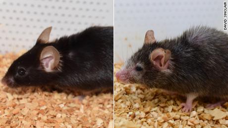
Stem cells are unspecialized cells that can be manipulated into becoming mature cells with special functions.
“Our mouse embryo model not only develops a brain, but also a beating heart, all the components that go on to make up the body,” said lead study author Magdalena Zernicka-Goetz, professor of mammalian development and stem cell biology at the University of Cambridge in the United Kingdom.
“It’s just unbelievable that we’ve got this far. This has been the dream of our community for years, and a major focus of our work for a decade, and finally we’ve done it.”
The paper is an exciting advance and tackles a challenge scientists face studying mammal embryos in utero, said Marianne Bronner, a professor of biology at the California Institute of Technology in Pasadena (Caltech). Bronner was not involved in the study.
“These develop outside of the mother and therefore can be easily visualized through critical developmental stages that were previously difficult to access,” Bronner added.
The researchers hope to move from mouse embryos to creating models of natural human pregnancies — many of which fail in the early stages, Zernicka-Goetz said.
By watching the embryos in a lab instead of a uterus, scientists got a better view into the process to learn why some pregnancies might fail and how to prevent it, she added.
For now, researchers have only been able to track about eight days of development in the mouse synthetic embryos, but the process is improving, and they are already learning a lot, said study author Gianluca Amadei, a postdoctoral researcher at the University of Cambridge.
“It reveals the fundamental requirements that have to be fulfilled to make the right structure of the embryo with its organs,” Zernicka-Goetz said.
Where it stands, the research doesn’t apply to humans and “there needs to be a high degree of improvement for this to be truly useful,” said Benoit Bruneau, the director of the Gladstone Institute of Cardiovascular Disease and a senior investigator at Gladstone Institutes. Bruneau was not involved in the study.
But researchers see important uses for the future. The process can be used immediately to test new drugs, Zernicka-Goetz said. But in the longer term, as scientists move from mouse synthetic embryos to a human embryo model, it also could help build synthetic organs for people who need transplants, Zernicka-Goetz added.
“I see this work as being the first example of work of this kind,” said study author David Glover, research professor of biology and biological engineering at Caltech.
How they did it
In utero, an embryo needs three types of stem cells to form: One becomes the body tissue, another the sac where the embryo develops, and the third the placenta connecting parent and fetus, according to the study.
In Zernicka-Goetz’s lab, researchers isolated the three types of stem cells from embryos and cultured them in a container angled to bring the cells together and encourage crosstalk between them.
Day by day, they were able to see the group of cells form into a more and more complex structure, she said.
There are ethical and legal considerations to address before moving to human synthetic embryos, Zernicka-Goetz said. And with the difference in complexity between mouse and human embryos, it could be decades before researchers are able to do a similar process for human models, Bronner said.
But in the meantime, the information learned from the mouse models could help “correct failing tissues and organs,” Zernicka-Goetz said.
The mystery of human life
The early weeks after fertilization are made up of these three different stem cells communicating with one another chemically and mechanically so the embryo can grow properly, the study said.
“So many pregnancies fail around this time, before most women (realize) they are pregnant,” said Zernicka-Goetz, who is also professor of biology and biological engineering at Caltech. “This period is the foundation for everything else that follows in pregnancy. If it goes wrong, the pregnancy will fail.”
But by this stage, an embryo created through in vitro fertilization is already implanted in the parent, so scientists have limited visibility into the processes it is going through, Zernicka-Goetz said.
They were able to develop foundations of a brain — a first for models such as these and a “holy grail for the field,” Glover said.
“This period of human life is so mysterious, so to be able to see how it happens in a dish — to have access to these individual stem cells, to understand why so many pregnancies fail and how we might be able to prevent that from happening — is quite special,” Zernicka-Goetz said in a press release. “We looked at the dialogue that has to happen between the different types of stem cell at that time — we’ve shown how it occurs and how it can go wrong.”
Summary: Researchers created model embryos from mouse stem cells that form a beating heart, a brain, and the foundation for other organs. The new model provides a novel way for future researchers to create and research the earliest stages of development.
Source: University of Cambridge
Researchers from the University of Cambridge have created model embryos from mouse stem cells that form a brain, a beating heart, and the foundations of all the other organs of the body – a new avenue for recreating the first stages of life.
The team, led by Professor Magdalena Zernicka-Goetz, developed the embryo model without eggs or sperm, and instead used stem cells – the body’s master cells, which can develop into almost any cell type in the body.
The researchers mimicked natural processes in the lab by guiding the three types of stem cells found in early mammalian development to the point where they start interacting. By inducing the expression of a particular set of genes and establishing a unique environment for their interactions, the researchers were able to get the stem cells to ‘talk’ to each other.
The stem cells self-organised into structures that progressed through the successive developmental stages until they had beating hearts and the foundations of the brain, as well as the yolk sac where the embryo develops and gets nutrients from in its first weeks. Unlike other synthetic embryos, the Cambridge-developed models reached the point where the entire brain, including the anterior portion, began to develop.
This is a further point in development than has been achieved in any other stem cell-derived model.
The team say their results, the result of more than a decade of research that progressively led to more and more complex embryo-like structures and reported today in the journal Nature, could help researchers understand why some embryos fail while others go on to develop into a healthy pregnancy. Additionally, the results could be used to guide repair and development of synthetic human organs for transplantation.
“Our mouse embryo model not only develops a brain, but also a beating heart, all the components that go on to make up the body,” said Zernicka-Goetz, Professor in Mammalian Development and Stem Cell Biology in Cambridge’s Department of Physiology, Development and Neuroscience, adding:
“It’s just unbelievable that we’ve got this far. This has been the dream of our community for years, and a major focus of our work for a decade, and finally we’ve done it.”
For a human embryo to develop successfully, there needs to be a ‘dialogue’ between the tissues that will become the embryo, and the tissues that will connect the embryo to the mother. In the first week after fertilisation, three types of stem cells develop: one will eventually become the tissues of the body, and the other two support the embryo’s development.
One of these extraembryonic stem cell types will become the placenta, which connects the foetus to the mother and provides oxygen and nutrients; and the second is the yolk sac, where the embryo grows and where it gets its nutrients from in early development.
Many pregnancies fail at the point when the three types of stem cells begin to send mechanical and chemical signals to each other, which tell the embryo how to develop properly.
“So many pregnancies fail around this time, before most women realise they are pregnant,” said Zernicka-Goetz, who is also Professor of Biology and Biological Engineering at Caltech. “This period is the foundation for everything else that follows in pregnancy. If it goes wrong, the pregnancy will fail.”
Over the past decade, Professor Zernicka-Goetz’s group in Cambridge has been studying these earliest stages of pregnancy, in order to understand why some pregnancies fail and some succeed.
“The stem cell embryo model is important because it gives us accessibility to the developing structure at a stage that is normally hidden from us due to the implantation of the tiny embryo into the mother’s womb.”
Zernicka-Goetz added: “This accessibility allows us to manipulate genes to understand their developmental roles in a model experimental system.”
To guide the development of their synthetic embryo, the researchers put together cultured stem cells representing each of the three types of tissue in the right proportions and environment to promote their growth and communication with each other, eventually self-assembling into an embryo.
The researchers found that the extraembryonic cells signal to embryonic cells by chemical signals but also mechanistically, or through touch, guiding the embryo’s development.
“This period of human life is so mysterious, so to be able to see how it happens in a dish – to have access to these individual stem cells, to understand why so many pregnancies fail and how we might be able to prevent that from happening – is quite special,” said Zernicka-Goetz.
“We looked at the dialogue that has to happen between the different types of stem cell at that time – we’ve shown how it occurs and how it can go wrong.”
A major advance in the study is the ability to generate the entire brain, in particular the anterior part, which has been a major goal in the development of synthetic embryos.
Natural (top) and synthetic (bottom) embryos side by side to show comparable brain and heart formation. Image credit: Amadei and Handford
This works in Zernicka-Goetz’s system because this part of the brain requires signals from one of the extraembryonic tissues to be able to develop. The team thought that this might be taking place from their 2018 and 2021 studies, which used the same component cells to develop into embryos at a slightly earlier stage.
Now, by pushing development just one day further, they can definitively say that their model is the very first to signal development of the anterior, and in fact the whole, brain.
“This opens new possibilities to study the mechanisms of neurodevelopment in an experimental model,” said Zernicka-Goetz.
“In fact, we demonstrate the proof of this principle in the paper by knocking out a gene already known to be essential for formation of the neural tube, precursor of the nervous system, and for brain and eye development. In the absence of this gene, the synthetic embryos show exactly the known defects in brain development as in an animal carrying this mutation.
“This means we can begin to apply this kind of approach to the many genes with unknown function in brain development.”
While the current research was carried out in mouse models, the researchers are developing similar human models with the potential to be directed towards the generation of specific organ types – to understand mechanisms behind crucial processes that would be otherwise impossible to study in real embryos. At present, UK law permits human embryos to be studied in the laboratory only up to the 14th day of development.
If the methods developed by Zernicka-Goetz’s team are shown to be successful with human stem cells in future, they could also be used to guide development of synthetic organs for patients awaiting transplants.
“There are so many people around the world who wait for years for organ transplants,” said Zernicka-Goetz. “What makes our work so exciting is that the knowledge coming out of it could be used to grow correct synthetic human organs to save lives that are currently lost. It should also be possible to affect and heal adult organs by using the knowledge we have on how they are made.
Professor Magdalena Zernicka-Goetz has made an incredible scientific breakthrough.
The creation of synthetic mouse embryos in a test tube that develop brains and beating hearts, starting only with embryonic stem cells, is the culmination of a decade’s work.
Magda explains: “I’m fascinated by the mystery of how embryos work. Every embryo follows a similar journey: one cell becomes many, then they communicate with each other and arrange themselves to form a structure that will provide a blueprint for all adult body parts. But how do embryo cells decide their fate, how do they know where to go and what to do? How do they form the right parts in the right place at the right time?
“Building the first ‘synthetic embryo’ models was a process we achieved step by step. Setting out, we knew that embryonic stem cells could be cultured indefinitely in the lab, and that when they’re injected into an embryo they can potentially contribute to any tissue in the adult organism. The challenge was to guide them to develop into a complete embryo.
“In addition to the embryonic stem cells we used two kinds of extraembryonic tissue: one of which forms the placenta and the other a sac in which the embryo develops. These tissues are very important, as they send signals to the embryo to develop all its parts at the right time and in the right place.
“Combining stem cells representing each of these three types of tissue is easier said than done. We had to find an environment where all three distinct cell types could grow and communicate with each other. And we had to find the right proportions of each cell type, and add them in the right sequence.
“Once we established these basic principles, the stem cells did the rest: they self-organised to progress through successive developmental stages until they had beating hearts and the foundations for a brain.
“The key to our achievement was thinking outside the conventional box. The majority of embryo model studies focus on embryonic stem cells, but don’t consider the significant role of extraembryonic cells. We mixed the right proportions of both embryonic and extraembryonic stem cells. Extraembryonic cells signal to embryonic cells through different means, by chemical signals but also mechanistically ‘by touch’. Our studies are helping to understand these signalling events.
“We are developing an analogous model of the human embryo, to understand the mechanisms behind crucial processes that would be otherwise impossible to study. This is important because the great majority of human pregnancies fail at this developmental stage, due to causes that we don’t understand. It will also allow us to identify factors permitting development of healthy human tissues as they form different organs.
“Creating the new ‘synthetic embryo’ has taught us a lot about the mechanisms by which the embryo builds itself. We learnt how the extraembryonic tissues direct the embryonic stem cells along the right pathways to signal formation of the correct structures; how cells move between compartments as the multi-layered body plan arises; and how this correctly sets the scene for neurulation – the process where tissue folds to form the neural tube and, in turn, the brain and spinal cord.
“This model gives us access to the developing structure at a stage that’s normally hidden from us, when the tiny embryo implants into the mother’s womb. Our model does not have to implant to develop, so it remains completely visible to us, allowing us to see the embryo’s progression through that developmental stage. This accessibility allows us to manipulate genes to understand their developmental roles in a model experimental system.
See also
“It’s certainly true that carrying out this type of work requires passion and resilience. I grew up inPoland under a Communist regime, which meant that traveling wasn’t allowed and thinking differently was not encouraged. There was immense social pressure to conform, and a lot of us rebelled against that. A silver lining of this was a desire to think independently and to persevere despite discouragement. That shaped me as a scientist too.
“When I started my research group in Cambridge, I established ways to study the ‘developmental black box’ – the development of the embryo at the time of implantation. My mentors had discouraged me from pursuing it during my PhD because they were concerned it would be difficult to shine light inside this ‘box.’ But I was so taken by the question of how the embryo self-organises that I didn’t give up and, inch by inch, we have worked our way forward.
“My advice to all young scientists is to follow your heart. Study a topic that inspires you, and choose an advisor who can be supportive of your style of work. In my opinion, it’s important to guide young scientists in the lab, but also to give them space to explore their individuality.
“My experience is that the challenges for female scientists increase as they progress through their careers. At later stages, it’s critical to have mentors who understand not only science but also how to balance it with everyday life, including starting a family.
“During my own pregnancy, I was shocked when an early screening showed abnormalities. The sampling was of extraembryonic cells so I waited for the amniocentesis, which samples foetal cells that have fallen into the amniotic fluid. These were normal, which put my mind at ease.
“The experience led me to study mosaic aneuploidy – a condition in which the embryo has cells with the wrong number of chromosomes alongside chromosomally normal cells. Incredibly, we found that these abnormal cells can be eliminated, and the normal, healthy cells compensate for their absence.
“For some reason this mechanism doesn’t operate in the tissues that build the placenta, and we’re still trying to understand why and how.
“Science is demanding, it’s hard work and it takes away most of your waking hours. I switch off by watching films – I watch a lot foreign films in Polish, French, and Danish, and documentaries and art films. But when I want to lose myself in another narrative, I watch dramas. I’m also a recent convert to gardening, where I can encourage the successful development of other life forms!
“It is an incredible feeling and a privilege to have this direct insight into the origins of a new life. It’s like discovering a new planet that we didn’t know existed.”
Funding: The research was supported in part by the European Research Council and Wellcome and NIH Pioneer Awards to Magdalena Zernicka-Goetz. Zernicka-Goetz is a Fellow of Sidney Sussex College, Cambridge.
About this genetics research news
Author: Sarah Collins and Jacqueline Garget Source: University of Cambridge Contact: Sarah Collins and Jacqueline Garget – University of Cambridge
Original Research: Closed access. “Synthetic embryos complete gastrulation to neurulation and organogenesis” by Magdalena Zernicka-Goetz et al. Nature
Abstract
Synthetic embryos complete gastrulation to neurulation and organogenesis
Embryonic stem cells (ESC) can undergo many aspects of mammalian embryogenesis in vitro, but their developmental potential is substantially extended by interactions with extraembryonic stem cells, including trophoblast stem cells (TSCs), extraembryonic endoderm stem cells (XEN), and inducible-XEN cells (iXEN).
Here, we assembled stem-cell derived embryos in vitro from mouse ESCs, TSCs and iXEN cells and showed that they recapitulate whole natural mouse embryo development in utero to day 8.5.
Our embryo model displays head-folds with defined forebrain and midbrain regions and develops a beating heart-like structure, a trunk comprising a neural tube and somites, a tail bud containing neuromesodermal progenitors, a gut tube, and primordial germ cells. This complete embryo model develops within an extra-embryonic yolk sac that initiates blood island development.
Importantly, we demonstrate that the neurulating embryo model assembled from Pax6 knockout-ESCs aggregated with wild-type TSCs and iXENs recapitulates the ventral domain expansion of the neural tube that occurs in natural, ubiquitous Pax6 knockout embryos. Thus, these complete embryoids are a powerful in vitro model for dissecting the roles of diverse lineages and genes in development.
Our results demonstrate the self-organization ability of embryonic and two types of extra-embryonic stem cells to reconstitute mammalian development through and beyond gastrulation to neurulation and early organogenesis.
Natural and synthetic embryos side by side show comparable brain and heart formation. Credit: Amadei and Handford
Scientists from the University of Cambridge have created model embryos from mouse stem cells that form a brain, a beating heart, and the foundations of all the other organs of the body. It represents a new avenue for recreating the first stages of life.
The team of researchers, led by Professor Magdalena Zernicka-Goetz, developed the embryo model without eggs or sperm. Instead, they used stem cells – the body’s master cells, which can develop into almost any cell type in the body.
“It’s just unbelievable that we’ve got this far. This has been the dream of our community for years, and major focus of our work for a decade and finally we’ve done it.” — Magdalena Zernicka-Goetz
By guiding the three types of stem cells found in early mammalian development to the point where they start interacting, the researchers mimicked natural processes in the lab. The scientists were able to get the stem cells to ‘talk’ to each other by inducing the expression of a particular set of genes and establishing a unique environment for their interactions.
The stem cells self-organized into structures that progressed through the successive developmental stages until they had beating hearts and the foundations of the brain They also had the yolk sac where the embryo develops and gets nutrients from in its first weeks. Unlike other synthetic embryos, the Cambridge-developed models reached the point where the entire brain, including the anterior portion, began to develop. This is a further point in development than has been achieved in any other stem cell-derived model.
According to the team, their results could help researchers understand why some embryos fail while others go on to develop into a healthy pregnancy. In addition, the results could be used to guide the repair and development of synthetic human organs for transplantation. The study, which is the result of more than a decade of research that progressively led to more and more complex embryo-like structures, was reported on August 25, 2022, in the journal Nature.
Natural and synthetic embryos side by side show comparable brain and heart formation. Credit: Amadei and Handford
“Our mouse embryo model not only develops a brain, but also a beating heart, all the components that go on to make up the body,” said Zernicka-Goetz, Professor in Mammalian Development and Stem Cell Biology in Cambridge’s Department of Physiology, Development and Neuroscience. “It’s just unbelievable that we’ve got this far. This has been the dream of our community for years, and major focus of our work for a decade and finally we’ve done it.”
A “dialog” between the tissues that will form the embryo and the tissues that will connect the embryo to the mother is necessary for the healthy development of a human embryo. Three different stem cell types begin to form in the first week following fertilization; one of these will eventually develop into the bodily tissues, while the other two support the embryo’s development. One of these extraembryonic stem cell types will become the placenta, which connects the fetus to the mother and provides oxygen and nutrients. The second is the yolk sac, where the embryo grows and where it gets its nutrients from in early development.
Many pregnancies fail at the point when the three types of stem cells begin to send mechanical and chemical signals to each other, which instruct the embryo on how to develop properly.
“So many pregnancies fail around this time, before most women realize they are pregnant,” said Zernicka-Goetz, who is also Professor of Biology and Biological Engineering at Caltech. “This period is the foundation for everything else that follows in pregnancy. If it goes wrong, the pregnancy will fail.”
Professor Zernicka-Goetz in the lab. Credit: University of Cambridge
Professor Zernicka-Goetz’s group in Cambridge has been studying these earliest stages of pregnancy over the past decade, in order to understand why some pregnancies fail and some succeed.
“The stem cell embryo model is important because it gives us accessibility to the developing structure at a stage that is normally hidden from us due to the implantation of the tiny embryo into the mother’s womb,” said Zernicka-Goetz. “This accessibility allows us to manipulate genes to understand their developmental roles in a model experimental system.”
To guide the development of their synthetic embryo, the scientists put together cultured stem cells representing each of the three types of tissue in the right proportions and environment to promote their growth and communication with each other, eventually self-assembling into an embryo.
The research team discovered that the extraembryonic cells signal to embryonic cells by chemical signals but also mechanistically, or through touch, guiding the embryo’s development.
“This period of human life is so mysterious, so to be able to see how it happens in a dish – to have access to these individual stem cells, to understand why so many pregnancies fail and how we might be able to prevent that from happening – is quite special,” said Zernicka-Goetz. “We looked at the dialogue that has to happen between the different types of stem cell at that time – we’ve shown how it occurs and how it can go wrong.”
A major advance in the study is the ability to generate the entire brain, in particular the anterior part, which has been a major goal in the development of synthetic embryos. This works in Zernicka-Goetz’s system because this part of the brain requires signals from one of the extraembryonic tissues to be able to develop. The team thought that this might be taking place from their 2018 and 2021 studies, which used the same component cells to develop into embryos at a slightly earlier stage. Now, by pushing development just one day further, they can definitively say that their model is the very first to signal development of the anterior, and in fact the whole, brain.
“This opens new possibilities to study the mechanisms of neurodevelopment in an experimental model,” said Zernicka-Goetz. “In fact, we demonstrate the proof of this principle in the paper by knocking out a gene already known to be essential for formation of the neural tube, precursor of the nervous system, and for brain and eye development. In the absence of this gene, the synthetic embryos show exactly the known defects in brain development as in an animal carrying this mutation. This means we can begin to apply this kind of approach to the many genes with unknown function in brain development.”
While the current research was carried out in mouse models, the researchers are developing similar human models with the potential to be directed towards the generation of specific organ types to understand mechanisms behind crucial processes that would be otherwise impossible to study in real embryos. At present, UK law permits human embryos to be studied in the laboratory only up to the 14th day of development.
If the methods developed by Zernicka-Goetz’s team are shown to be successful with human stem cells in the future, they could also be used to guide development of synthetic organs for patients awaiting transplants.
“There are so many people around the world who wait for years for organ transplants,” said Zernicka-Goetz. “What makes our work so exciting is that the knowledge coming out of it could be used to grow correct synthetic human organs to save lives that are currently lost. It should also be possible to affect and heal adult organs by using the knowledge we have on how they are made.
“This is an incredible step forward and took 10 years of hard work of many of my team members – I never thought we’d get to this place. You never think your dreams will come true, but they have.”
Professor Magdalena Zernicka-Goetz has made an incredible scientific breakthrough.
The creation of synthetic mouse embryos in a test tube that develop brains and beating hearts, starting only with embryonic stem cells, is the culmination of a decade’s work.
Magda explains:
I’m fascinated by the mystery of how embryos work. Every embryo follows a similar journey: one cell becomes many, then they communicate with each other and arrange themselves to form a structure that will provide a blueprint for all adult body parts. But how do embryo cells decide their fate, how do they know where to go and what to do? How do they form the right parts in the right place at the right time?
Building the first ‘synthetic embryo’ models was a process we achieved step by step. Setting out, we knew that embryonic stem cells could be cultured indefinitely in the lab, and that when they’re injected into an embryo they can potentially contribute to any tissue in the adult organism. The challenge was to guide them to develop into a complete embryo. In addition to the embryonic stem cells we used two kinds of extraembryonic tissue: one of which forms the placenta and the other a sac in which the embryo develops. These tissues are very important, as they send signals to the embryo to develop all its parts at the right time and in the right place.
Combining stem cells representing each of these three types of tissue is easier said than done. We had to find an environment where all three distinct cell types could grow and communicate with each other. And we had to find the right proportions of each cell type, and add them in the right sequence. Once we established these basic principles, the stem cells did the rest: they self-organized to progress through successive developmental stages until they had beating hearts and the foundations for a brain.
The key to our achievement was thinking outside the conventional box. The majority of embryo model studies focus on embryonic stem cells, but don’t consider the significant role of extraembryonic cells. We mixed the right proportions of both embryonic and extraembryonic stem cells. Extraembryonic cells signal to embryonic cells through different means, by chemical signals but also mechanistically ‘by touch’. Our studies are helping to understand these signaling events.
We are developing an analogous model of the human embryo, to understand the mechanisms behind crucial processes that would be otherwise impossible to study. This is important because the great majority of human pregnancies fail at this developmental stage, due to causes that we don’t understand. It will also allow us to identify factors permitting development of healthy human tissues as they form different organs.
Creating the new ‘synthetic embryo’ has taught us a lot about the mechanisms by which the embryo builds itself. We learned how the extraembryonic tissues direct the embryonic stem cells along the right pathways to signal formation of the correct structures; how cells move between compartments as the multi-layered body plan arises; and how this correctly sets the scene for neurulation — the process where tissue folds to form the neural tube and, in turn, the brain and spinal cord.
This model gives us access to the developing structure at a stage that’s normally hidden from us, when the tiny embryo implants into the mother’s womb. Our model does not have to implant to develop, so it remains completely visible to us, allowing us to see the embryo’s progression through that developmental stage. This accessibility allows us to manipulate genes to understand their developmental roles in a model experimental system.
It’s certainly true that carrying out this type of work requires passion and resilience. I grew up in Poland under a Communist regime, which meant that traveling wasn’t allowed and thinking differently was not encouraged. There was immense social pressure to conform, and a lot of us rebelled against that. A silver lining of this was a desire to think independently and to persevere despite discouragement. That shaped me as a scientist too.
When I started my research group in Cambridge, I established ways to study the ‘developmental black box’ — the development of the embryo at the time of implantation. My mentors had discouraged me from pursuing it during my PhD because they were concerned it would be difficult to shine light inside this ‘box.’ But I was so taken by the question of how the embryo self-organizes that I didn’t give up and, inch by inch, we have worked our way forward.
My advice to all young scientists is to follow your heart. Study a topic that inspires you, and choose an advisor who can be supportive of your style of work. In my opinion, it’s important to guide young scientists in the lab, but also to give them space to explore their individuality. My experience is that the challenges for female scientists increase as they progress through their careers. At later stages, it’s critical to have mentors who understand not only science but also how to balance it with everyday life, including starting a family.
During my own pregnancy, I was shocked when an early screening showed abnormalities. The sampling was of extraembryonic cells so I waited for the amniocentesis, which samples fetal cells that have fallen into the amniotic fluid. These were normal, which put my mind at ease. The experience led me to study mosaic aneuploidy — a condition in which the embryo has cells with the wrong number of chromosomes alongside chromosomally normal cells. Incredibly, we found that these abnormal cells can be eliminated, and the normal, healthy cells compensate for their absence. For some reason, this mechanism doesn’t operate in the tissues that build the placenta, and we’re still trying to understand why and how.
Science is demanding, it’s hard work and it takes away most of your waking hours. I switch off by watching films — I watch a lot of foreign films in Polish, French, and Danish, and documentaries and art films. But when I want to lose myself in another narrative, I watch dramas. I’m also a recent convert to gardening, where I can encourage the successful development of other life forms!
It is an incredible feeling and a privilege to have this direct insight into the origins of a new life. It’s like discovering a new planet that we didn’t know existed.
For more on this research, see Scientists Grow “Synthetic” Mouse Embryo With Brain and Beating Heart.
Reference: “Synthetic embryos complete gastrulation to neurulation and organogenesis” by Gianluca Amadei, Charlotte E. Handford, Chengxiang Qiu, Joachim De Jonghe, Hannah Greenfeld, Martin Tran, Beth K. Martin, Dong-Yuan Chen, Alejandro Aguilera-Castrejon, Jacob H. Hanna, Michael Elowitz, Florian Hollfelder, Jay Shendure, David M. Glover and Magdalena Zernicka-Goetz, 25 August 2022, Nature. DOI: 10.1038/s41586-022-05246-3
Scientists have created the world’s first lab-grown “synthetic embryo,” a groundbreaking moment in science that has reignited a fierce ethical debate.
Led by molecular geneticist Joseph Hanna, a team of researchers at Israel’s Wizemann Institute of Science managed to create a synthetic mouse “embryo” in a lab without fertilized eggs or a uterus, potentially allowing us to get a glimpse of what happens in the early stages of human pregnancy as well.
This new embryo model, as detailed in the team’s paper published this week in the journal Cell, was able to mimic all the makings of an early body, “including precursors of heart, blood, brain and other organs” as well as “the ‘support’ cells like those found in the placenta and other tissues required to establish and maintain a pregnancy,” as University of Melbourne stem cell researcher Megan Munsie, who was not involved in the research, wrote in a piece for The Conversation.
The research could have major implications.
“This is a crucial stage: in humans, many pregnancies are lost around this stage, and we don’t really know why,” Munsie wrote. “Having models provides a way to better understand what can go wrong, and possibly insights into what we may be able to do about it.”
The embryo model, however, only survived eight out of the 20-day mouse embryonic cycle, a critical drawback that, given the stated goal of Renewal Bio, the company founded by Hanna to commercially fund this research.
The startup’s goal is to develop synthetic human stem cells in an attempt to “solve” human health crises, a science which experts say won’t be ready for decades.
In short, Bio Renewal wants to create embryo-stage versions of humans so that it can harvest tissues for transplants.
Critics who spoke with MIT Technology Review said that it wasn’t time to talk about the creation of synthetic human embryos, especially given the greater political context and controversy surrounding the research.
“It’s absolutely not necessary,” Nicolas Rivron, a stem-cell scientist at Vienna’s Institute of Molecular Biotechnology, told the magazine, “so why would you do it?”
He’s not alone in his criticisms, either.
“Synthetic human embryos are not an immediate prospect,” James Briscoe of the Francis Crick Institute in London told The Guardian in response to the new research.
“We know less about human embryos than mouse embryos and the inefficiency of the mouse synthetic embryos suggests that translating the findings to human requires further development,” he added.
It seems that regardless of where researchers fall on the topic, most agree that it’s way too early to start seriously talking about the ethics of synthetic human embryos — but it’s a breakthrough nonetheless.
READ MORE: This startup wants to copy you into an embryo for organ harvesting [MIT Technology Review]
More on stem cell promises: Scientist Says Humans Will Be Able to Regrow Limbs “Within Our Lifetimes”
Without Egg, Sperm or Womb: Synthetic Mouse Embryo Models Created Solely from Stem Cells
An egg meets a sperm – that’s a necessary first step in life’s beginnings. In embryonic development research, it’s also a common first step. However, in a new study published on August 1, 2022, in the journal Cell, researchers from the Weizmann Institute of Science have grown synthetic embryo models of mice outside the womb by starting solely with stem cells cultured in a petri dish. That means they are grown without the use of fertilized eggs. This method opens new horizons for studying how stem cells form various organs in the developing embryo. It may also one day make it possible to grow tissues and organs for transplantation using synthetic embryo models.
A video showing a synthetic mouse embryo model on day 8 of its development; it has a beating heart, a yolk sac, a placenta, and emerging blood circulation.
“The embryo is the best organ-making machine and the best 3D bioprinter – we tried to emulate what it does,” says Prof. Jacob Hanna of Weizmann’s Molecular Genetics Department, who headed the research team.
Hanna explains that scientists already know how to restore mature cells to “stemness.” In fact, pioneers of this cellular reprogramming won a Nobel Prize in 2012. However, going in the opposite direction, that is, causing stem cells to differentiate into specialized body cells, not to mention form entire organs, has proved far more difficult.
“Until now, in most studies, the specialized cells were often either hard to produce or aberrant, and they tended to form a mishmash instead of well-structured tissue suitable for transplantation. We managed to overcome these hurdles by unleashing the self-organization potential encoded in the stem cells.”
(Left to right): Dr. Noa Novershtern, Prof. Jacob Hanna, Alejandro Aguilera-Castrejon, Shadi Tarazi and Carine Joubran. Credit: Weizmann Institute of Science
Hanna’s team built on two previous advances in his lab. One was an efficient method for reprogramming stem cells back to a naïve state – that is, to their earliest stage – when they have the greatest potential to specialize into different cell types. The other, described in a scientific paper in Nature in March 2021, was the electronically controlled device the team had developed over seven years of trial and error for growing natural mouse embryos outside the womb. The device keeps the embryos bathed in a nutrient solution inside of beakers that move continuously, simulating the way nutrients are supplied by material blood flow to the placenta, and closely controls oxygen exchange and atmospheric pressure. In the earlier research, the team had successfully used this device to grow natural mouse embryos from day 5 to day 11.
This is how synthetic mouse embryo models were grown outside the womb: a video showing the device in action. Continuously moving beakers simulate the natural nutrient supply, while oxygen exchange and atmospheric pressure are tightly controlled.
In the new study, the team set out to grow a synthetic embryo model solely from naïve mouse stem cells that had been cultured for years in a petri dish, dispensing with the need for starting with a fertilized egg. This approach is extremely valuable because it could, to a large extent, bypass the technical and ethical issues involved in the use of natural embryos in research and biotechnology. Even in the case of mice, certain experiments are currently unfeasible because they would require thousands of embryos, whereas access to models derived from mouse embryonic cells, which grow in lab incubators by the millions, is virtually unlimited.
“The embryo is the best organ-making machine and the best 3D bioprinter – we tried to emulate what it does.”
Before placing the stem cells into the device, the researchers separated them into three groups. In one, which contained cells intended to develop into embryonic organs themselves, the cells were left as they were. Cells in the other two groups were pretreated for only 48 hours to overexpress one of two types of genes: master regulators of either the placenta or the yolk sac. “We gave these two groups of cells a transient push to give rise to extraembryonic tissues that sustain the developing embryo,” Hanna says.
Development of synthetic embryo models from day 1 (top left) to day 8 (bottom right). All their early organ progenitors had formed, including a beating heart, an emerging blood circulation, a brain, a neural tube, and an intestinal tract. Credit: Weizmann Institute of Science
Soon after being mixed together inside the device, the three groups of cells convened into aggregates, the vast majority of which failed to develop properly. But about 0.5 percent – 50 of around 10,000 – went on to form spheres, each of which later became an elongated, embryo-like structure. Since the researchers had labeled each group of cells with a different color, they were able to observe the placenta and yolk sacs forming outside the embryos and the model’s development proceeding as in a natural embryo. These synthetic models developed normally until day 8.5 – nearly half of the mouse 20-day gestation – at which stage all the early organ progenitors had formed, including a beating heart, blood stem cell circulation, a brain with well-shaped folds, a neural tube and an intestinal tract. When compared to natural mouse embryos, the synthetic models displayed a 95 percent similarity in both the shape of internal structures and the gene expression patterns of different cell types. The organs seen in the models gave every indication of being functional.
Day 8 in the life of a mouse embryo: a synthetic model (top) and a natural embryo (bottom). The synthetic models displayed a 95 percent similarity in both the shape of internal structures and the gene expression patterns of different cell types. Credit: Weizmann Institute of Science
For Hanna and other stem cell and embryonic development researchers, the study presents a new arena: “Our next challenge is to understand how stem cells know what to do – how they self-assemble into organs and find their way to their assigned spots inside an embryo. And because our system, unlike a womb, is transparent, it may prove useful for modeling birth and implantation defects of human embryos.”
In addition to helping reduce the use of animals in research, synthetic embryo models might in the future become a reliable source of cells, tissues, and organs for transplantation. “Instead of developing a different protocol for growing each cell type – for example, those of the kidney or liver – we may one day be able to create a synthetic embryo-like model and then isolate the cells we need. We won’t need to dictate to the emerging organs how they must develop. The embryo itself does this best.”
A diagram showing the innovative method for growing synthetic mouse embryo models from stem cells – without egg, sperm or womb – developed in the laboratory of Prof. Jacob Hanna. Credit: Weizmann Institute of Science
Reference: “Post-Gastrulation Synthetic Embryos Generated Ex Utero from Mouse Naïve ESCs” by Shadi Tarazi, Alejandro Aguilera-Castrejon, Carine Joubran, Nadir Ghanem, Shahd Ashouokhi, Francesco Roncato, Emilie Wildschutz, Montaser Haddad, Bernardo Oldak, Elidet Gomez-Cesar, Nir Livnat, Sergey Viukov, Dmitry Lukshtanov, Segev Naveh-Tassa, Max Rose, Suhair Hanna, Calanit Raanan, Ori Brenner, Merav Kedmi, Hadas Keren-Shaul, Tsvee Lapidot, Itay Maza, Noa Novershtern and Jacob H. Hanna, 1 August 2022, Cell. DOI: 10.1016/j.cell.2022.07.028
This research was co-led by Shadi Tarazi, Alejandro Aguilera-Castrejon, and Carine Joubran of Weizmann’s Molecular Genetics Department. Study participants also included Shahd Ashouokhi, Dr. Francesco Roncato, Emilie Wildschutz, Dr. Bernardo Oldak, Elidet Gomez-Cesar, Nir Livnat, Sergey Viukov, Dmitry Lokshtanov, Segev Naveh-Tassa, Max Rose and Dr. Noa Novershtern of Weizmann’s Molecular Genetics Department; Montaser Haddad and Prof. Tsvee Lapidot of Weizmann’s Immunology and Regenerative Biology Department; Dr. Merav Kedmi of Weizmann’s Life Sciences Core Facilities Department; Dr. Hadas Keren-Shaul of the Nancy and Stephen Grand Israel National Center for Personalized Medicine; and Dr. Nadir Ghanem, Dr. Suhair Hanna and Dr. Itay Maza of the Rambam Health Care Campus.
Prof. Jacob Hanna’s research is supported by the Dr. Barry Sherman Institute for Medicinal Chemistry; the Helen and Martin Kimmel Institute for Stem Cell Research; and Pascal and Ilana Mantoux.
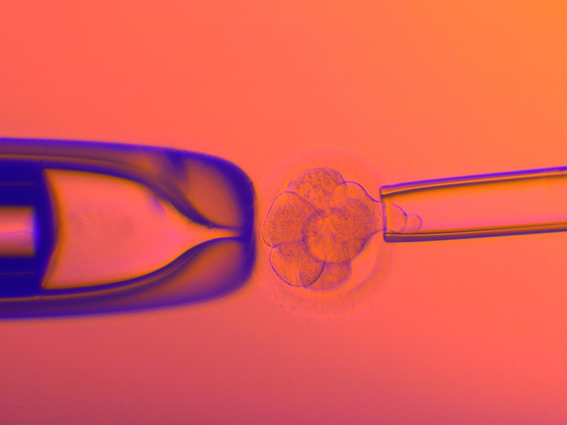
A cell is plucked from a human embryo created using in vitro fertilization so that it can be screened for genetic disorders.Credit: Pascal Goetgheluck/SPL
The emergence of companies that offer prospective parents complex genetic tests on embryos ahead of in vitro fertilization (IVF) has alarmed geneticists and bioethicists alike. The companies claim to be able to predict the risk of many common diseases — including those influenced by dozens or even hundreds of genes. People undergoing IVF are then offered the chance to select an embryo with a perceived low relative risk of developing such diseases.
Researchers are right to be concerned. The selection of embryos on the basis of these predictions is not yet supported by science. Moreover, the societal implications of using complex genetic tests to choose embryos has not yet been fully considered. Some scientists are completely opposed to the practice, whereas others recognize that, as more data accrue, there might be benefits, but realize that it must be carefully regulated. A study published in Nature Medicine1 on 21 March that explains some of the methodology behind the determination of what are called polygenic risk scores draws attention to the practice — but does not allay scientists’ fears.
Some health authorities around the world do regulate the use of simple genetic testing alongside IVF, but many do not. The aim of these tests is to reduce the chances of a parent transmitting an inherited disease to their unborn child. This is typically done in the case of rare, devastating conditions caused by mutations in a single gene. In the United Kingdom, for example, tests have been approved by the Human Fertilisation and Embryology Authority for more than 600 inherited disorders, including Tay–Sachs disease and breast cancers caused by mutations in the genes BRCA1 and BRCA2.
But the most common diseases, such as type 2 diabetes, are associated with mutations not in a single gene, but in many — potentially even thousands. To understand genetic contributions to such conditions, researchers have been analysing DNA sequences from many thousands of people with the disease, and comparing them with the DNA of people who do not have it, looking for genetic variants that are associated with a higher risk of developing the condition. This information is then converted into an overall risk score that estimates a person’s relative risk of developing a given disorder.
The ambition is that, as genetic studies continue to sample populations more broadly and more deeply, polygenic risk scores will become more refined, and could eventually be used to guide treatment and prevention strategies. But there’s a consensus that the scores are not yet ready to be used beyond research studies.
Feasibility study
In the latest study, the authors — most of whom work for companies involved in IVF or genetic testing — address the technical challenge of predicting accurate genome sequences from the small amounts of DNA available from one or two cells biopsied from an embryo. The researchers constructed sequences for more than 100 embryos by analysing hundreds of thousands of sites in the embryos’ genomes using a technique called genotyping, which requires less DNA than whole-genome sequencing does. They then combined these data with whole-genome sequences from the relevant prospective parents to fill in the rest of the DNA sequences and compared the reconstructed genomes with those of ten born siblings. They found that they were able to infer the correct genome sequence at sites used to calculate polygenic risk scores for 12 conditions — including diabetes, certain types of heart disease and several forms of cancer and autoimmune disorder — with 97–99% accuracy.
The authors say that this technique — which has been peer reviewed — establishes the feasibility of assessing genomic regions necessary to calculate a polygenic risk score for an embryo. But this technical capability is not the main reason for concern and debate over the use of polygenic risk scores in embryo selection for IVF.
There are many other concerns surrounding the practice. One is that the scores have been developed on the basis of genome-wide association studies that have heavily sampled DNA from people of European descent. Although efforts are under way to diversify such databases, the scores currently available are not based on an appropriately diverse subset of people. Even among white European people, polygenic risk scores are sometimes predictive only within narrow subsets of that population — potentially, in part, because of poorly understood interactions between the genetic and environmental contributions to a condition.
Furthermore, scientists do not yet fully understand how the selection of embryos with a lower relative risk for one disease might influence susceptibility to other conditions. Genetic variation can have a number of effects — a phenomenon known as pleiotropy — and a DNA sequence associated with one beneficial characteristic could also increase the risk of a detrimental one.
Many of these polygenic scores are being used to predict the risk of disorders that occur later in life, without any way of incorporating potential changes in environment that could occur over that time. A child born today will probably not experience heart disease or diabetes for decades, and there is no way of knowing what treatments or preventive steps will be available by then, or what changes in the environment might have taken place.
Potential harms
Concerns about under-represented populations and pleiotropy might be addressed with further research. But polygenic risk assessments are already being marketed directly to consumers (and not only for IVF) in some countries, including the United States and Japan. It is not clear to what extent individuals are counselled on the technique’s uncertainties and risks. Meanwhile, the scores of such assessments could be harmful. They could trigger the unnecessary destruction of viable embryos or induce women to undergo extra cycles of ovarian stimulation to collect more oocytes.
In some regions, there is little regulation of IVF and a long history of IVF technologies sweeping the market with little evidence that they improve the chances of conception or offer health benefits to children born as a result of the technology. Meanwhile, IVF is big business: the global market was about US$14 billion in 2020 and is predicted to more than double, to $34 billion, by 2028, according to Grand View Research, a market-research company based in San Francisco, California.
For now, prospective parents seeking IVF should not be offered polygenic risk scores for diseases unless they are part of rigorous clinical trials. Professional societies should make this clear to their members — as some have already done — and should publish guidelines on how to counsel participants in such trials to avoid giving them false hopes or fears about their children’s health. Genetic counsellors must be trained to do the same.
These tests demand a broader societal discussion. By nature of their complexity, polygenic risk scores also open the door to evaluating not only disease risk, but traits such as height or intelligence. At present, not enough is known about the genetic contributors to such traits to develop meaningful tests that would allow prospective parents to select embryos. But those data are on the way and the technology is going to move quickly — it is well past time to discuss how far it should go.
A California company says it can decipher almost all the DNA code of a days-old embryo created through in vitro fertilization (IVF)—a challenging feat because of the tiny volume of genetic material available for analysis. The advance depends on fully sequencing both parents’ DNA and “reconstructing” an embryo’s genome with the help of those data. And the company suggests it could make it possible to forecast risk for common diseases that develop decades down the line. Currently, such genetic risk prediction is being tested in adults, and sometimes offered clinically. The idea of applying it to IVF embryos has generated intense scientific and ethical controversy. But that hasn’t stopped the technology from galloping ahead.
Heart conditions, autoimmune diseases, cancer, and many other adult ailments have complex and often mysterious origins, fueled by a mix of genetic and environmental influences. Hundreds of variations in the human genome can collectively raise or lower risk of a particular disease, sometimes by a lot. Predicting a person’s chance of a specific illness by blending this genetic variability into what’s called a “polygenic risk score” remains under study in adults, in part because our understanding of how gene variants come together to drive or protect against disease remains a work in progress. In embryos it’s even harder to prove a risk score’s accuracy, researchers say. “Ultimately, how are we going to validate this in embryos?” says Norbert Gleicher, an infertility specialist at the Center for Human Reproduction in New York City who was not involved in the research. “We’ll have to wait for 40 or 50 years” to find out whether a person develops the diseases they were screened for as an embryo.
With current technologies, it’s very difficult to accurately sequence a whole genome from just a few cells, though some have tried with different methods. The new work on polygenic risk scores for IVF embryos is “exploratory research,” says Premal Shah, CEO of MyOme, the company reporting the results. Today in Nature Medicine, the MyOme team, led by company co-founders and scientists Matthew Rabinowitz and Akash Kumar, along with colleagues elsewhere, describe creating such scores by first sequencing the genomes of 10 pairs of parents who had already undergone IVF and had babies. The researchers then used data collected during the IVF process: The couples’ embryos, 110 in all, had undergone limited genetic testing at that time, a sort of spot sequencing of cells, called microarray measurements. Such analysis can test for an abnormal number of chromosomes, certain genetic diseases, and rearrangements of large chunks of DNA, and it has become an increasingly common part of IVF treatment in the United States. By combining these patchy embryo data with the more complete parental genome sequences, and applying statistical and population genomics techniques, the researchers could account for the gene shuffling that occurs during reproduction and calculate which chromosomes each parent had passed down to each embryo. In this way, they could predict much of that embryo’s DNA.
The researchers had a handy way to see whether their reconstruction was accurate: Check the couples’ babies. They collected cheek swab samples from the babies and sequenced their full genome, just as they’d done with the parents. They then compared that “true sequence” with the reconstructed genome for the embryo from which the child originated. The comparison revealed, essentially, a match: For a 3-day-old embryo, at least 96% of the reconstructed genome aligned with the inherited gene variants in the corresponding baby; for a 5-day-old embryo, it was at least 98%. (Because much of the human genome is the same across all people, the researchers focused on the DNA variability that made the parents, and their babies, unique.)
“What they presented is a nice method to sequence the genomes of all embryos,” says Shai Carmi, a statistical geneticist at the Hebrew University of Jerusalem. Such an accomplishment “is not trivial.” Kumar hopes being able to reconstruct most of an embryo’s genome will provide information well beyond what’s now available to people undergoing IVF, to determine an offspring’s chances of staying healthy. “It’s not enough to focus on the single gene anymore,” he says.
Once they had reconstructed embryo genomes in hand, the researchers turned to published data from large genomic studies of adults with or without common chronic diseases and the polygenic risk score models that were derived from that information. Then, MyOme applied those models to the embryos, crunching polygenic risk scores for 12 diseases, including breast cancer, coronary artery disease, and type 2 diabetes. The team also experimented with combining the reconstructed embryo sequence of single genes, such as BRCA1 and BRCA2, that are known to dramatically raise risk of certain diseases, with an embryo’s polygenic risk scores for that condition—in this case, breast cancer.
“We’re talking about providing information on risks that people care about—heart disease, cancer, autoimmune disease,” says Kumar, who is also a pediatric medical geneticist. He still sees patients and sometimes encounters frustration from parents wanting to avoid conferring a high risk of ailments that run in their families to their offspring. At the same time, Kumar stresses, “This is a new technology. It’s going to have controversies and challenges.”
In fact, many researchers say it’s premature to use polygenic risk scores to select which embryos are transferred. Such risk scores are “primarily still a research tool, even in adults,” says Barbara Koenig, a medical anthropologist who works on bioethics at the University of California, San Francisco. She’s involved in a large study called Women Informed to Screen Depending On Measures of risk that offers some women polygenic risk scores for breast cancer along with screening recommendations. “The scores are constantly being refined, every week they change,” Koenig says. “It’s like a constantly moving target.”
Kumar and his co-authors acknowledge the scores’ limitations, including that they are based on DNA from populations of overwhelmingly European ancestry and may be less accurate in other groups. Because of that, the MyOme team did not create disease risk assessments for embryos whose genome reflected at least 20% Asian or African ancestry. Even the DNA array technologies used to reconstruct the embryonic genomes have a European bias, says Genevieve Wojcik, a genetic epidemiologist at Johns Hopkins University, and may be less reliable for those with non-European ancestry. “You have a tool that cannot be used for a large proportion of the population,” she says. Kumar says the company is working to make the technology more broadly applicable.
There are other concerns, too. Although Carmi says the accuracy of polygenic risk scores in adults has improved, it’s unknown whether scores based on adult DNA and health data translate to embryos, in part because the environment can play a major role in shaping outcomes. “It’s difficult to say whether this will be meaningful,” Carmi says. He and his colleagues have seen this limitation up close: They’ve used computer modeling to assess whether height and IQ can be boosted by selecting embryos using polygenic risk scores for either trait, and found that generally, it doesn’t work. “We’re still missing a lot” when it comes to understanding genetics, even for highly heritable traits such as height, he says. In another computer modeling paper, however, Carmi found certain disease polygenic risk scores in embryos may prove useful. That’s because unlike height, which runs across a spectrum, heart attacks, say, either happen or they don’t. And pulling down genetic risk somewhat by implanting a different embryo, he says, may be enough to avoid that outcome.
But like a painting with only one corner visible, much of the human genome remains shrouded, including how genes interact with each other and the multiple effects one gene may have. Gleicher worries about the unintended consequences of applying polygenic risk scores to embryos. “You can achieve omission of one disease but at the same time, by doing that, induce another disease.” For example, modeling suggests selecting an embryo with a high polygenic risk score for educational attainment could also increase its risk for bipolar disorder. In December 2021, the European Society of Human Genetics urged against using polygenic risk scores for embryo selection—a position firmly endorsed by Gleicher, who calls such practice “unethical.”
Still, some companies and fertility clinics already claim they can help parents select embryos for IQ and risk of various diseases. MyOme, meanwhile, is applying the methods from this latest study to another that’s ongoing, working with IVF clinics and couples who want to learn polygenic risk scores for their frozen embryos. Couples may opt to decide which embryos to implant based on that information. “When you have a lot of information presented in this context, is it going to provide empowerment, or is it just going to confuse the parents?” Kumar asks. That’s one question he hopes this ongoing study can answer.
Kumar says he’s well aware of the criticisms, including that polygenic risk scores may not even be accurate for embryos. “That point is heard,” Kumar says. “Our focus is doing this research because we see promise.”
Life reconstruction of a close-to-hatching oviraptorosaur dinosaur embryo, based on the new specimen “Baby Yingliang.” Credit: Lida Xing
Over the last 100 years, many fossilized dinosaur eggs and nests have been found, but finding one with a well-preserved embryo inside is exceedingly rare. Now, researchers reporting in the journal iScience on December 21, 2021, have detailed one such specimen discovered in southern China.
What’s more, their studies lead them to suggest that oviraptorosaurs (a group of therapods closely related to birds) took on a distinctive tucking posture before they hatched, a behavior that had been considered unique to birds. It raises the possibility that tucking behavior may have evolved first among non-avian theropods during the Animated life reconstruction of a close-to-hatching oviraptorosaur dinosaur embryo, based on the new specimen “Baby Yingliang.” Credit: Lida Xing
It had been acquired in 2000 by Liang Liu, director of a company called Yingliang Group, who suspected it might contain egg fossils. But it then ended up in storage, largely forgotten until about ten years later, when museum staff during the construction of Yingliang Stone Nature History Museum sorted through the boxes and unearthed the fossils.
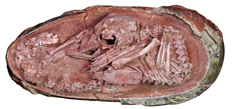
“Museum staff identified them as dinosaur eggs and saw some bones on the broken cross section of one of the eggs,” Lida Xing of China University of Geosciences, Beijing, said. The fossils were then prepared, unveiling the embryo hidden within, which they named “Baby Yingliang.”
Photo of the oviraptorosaur embryo “Baby Yingliang.” Credit: Xing et al./iScience
In the new study, Xing and colleagues report that the head lies ventral to the body, with the feet on either side, and the back curled along the blunt pole of the egg, in a posture previously unrecognized in a non-avian dinosaur. That’s especially notable because it’s reminiscent of a late-stage modern bird embryo.
Comparison of the specimen to other late-stage oviraptorosaur embryos suggests that before hatching, oviraptorosaurs developed avian-like postures late in their incubation. In modern birds, such coordinated embryonic movements are associated with tucking, a behavior that’s controlled by the central nervous system and is critical for hatching success.
The notion that such pre-hatching behavior may have originated among non-avian theropods can now be further investigated through more studies of other fossil embryos. But first, the researchers say they’ll continue studying this rare specimen in even more depth, using various imaging techniques to image its internal anatomy, such as skull bones, and other body parts that are still covered in rocks.
For more on this discovery, read Exquisitely Preserved Dinosaur Embryo Found Inside Fossilized Oviraptorosaur Egg.
Reference: “An exquisitely preserved in-ovo theropod dinosaur embryo sheds light on avian-like prehatching postures” by Lida Xing, Kecheng Niu, Waisum Ma, Darla K. Zelenitsky, Tzu-Ruei Yang and Stephen L. Brusatte, 21 December 2021, iScience. DOI: 10.1016/j.isci.2021.103516
This work was supported by the National Natural Science Foundation of China, 111 Project, Fundamental Research Funds for the Central Universities, the Natural Sciences and Engineering Research Council of Canada, and the Ministry of Science and Technology, Taiwan.
An exquisitely preserved dinosaur embryo was discovered inside a fossilized egg in China dating back 72 million years and is one of the most complete dinosaur embryos ever found. The amazing discovery helps prove that feather and flight evolution began in dinosaurs
Birds, descended from feathered theropod dinosaurs, are the only known living dinosaurs. The fascinating evolutionary mechanism of flight started in the Cretaceous period, an extinction event period that occurred 66 million years, which caused a sudden mass extinction of close to 75% of the earth’s floral and faunal species. Pertaining to this very subject, a fascinating new study has been published in iScience magazine, which has analyzed the so-called “Baby Yingliang” dinosaur embryo, a fossilized dinosaur egg, to further support the notion that bird evolution began with a certain kind of dinosaur.
“This dinosaur embryo inside its egg is one of the most beautiful fossils I have ever seen,” said paper co-author and vertebrate paleontologist Steve Brusatte of the University of Edinburgh. “This little prenatal dinosaur looks just like a baby bird curled in its egg, which is yet more evidence that many features characteristic of today’s birds first evolved in their dinosaur ancestors.”
Based on Baby Yingliang’s Dinosaur Embryo
Dated to between 72 and 66 million years ago (or mya), the dinosaur embryo dubbed “Baby Yingliang” was discovered in the late Cretaceous rocks of Ganzhou in southern China, reports Archaeology News Network . The species, known as oviraptorosaurs, are toothless, beaked, theropod dinosaurs. The dinosaur embryo was discovered in the year 2000 and housed in the Yingliang Stone Natural History Museum in Xiamen, Fujian, China. The current study was led by paleontologists from the University of Birmingham, UK, China’s University of Geosciences (Beijing), and other researchers from China, the UK, and Canada.
This find is one of the most complete dinosaur embryos ever found. The posture of the dinosaur in the embryo fossil prior to hatching matches that of birds. The scientists estimate the dinosaur would have been about 10.6 inches (27 centimeters) long at birth. The egg itself is 6.7 inches (17 centimeters) long. Bruscatte added that this egg is “more evidence that many features of today’s birds first evolved in their dinosaur ancestors.”
The Baby Yingliang dinosaur embryo used in the latest study to show that dinosaurs first developed feathers and then flight. Source: iScience
Pre-Hatching Behavior: Link Between Past and Present?
In the Baby Yingliang dinosaur embryo the head lies below the body, with the feet on either side, and the back curled along the blunt end of the egg. This phenomenon, witnessed in modern birds , is called tucking. This posture is seen critical to hatching, and thus controlled by the central nervous system . This has led scientists from the current study to suggest that this pre-hatching behavior may have originated first amongst non-avian theropods.
“It is interesting to see this dinosaur embryo and a chicken embryo pose in a similar way inside the egg, which possibly indicates similar prehatching behaviors,” said Fion Waisum Ma, joint first author and PhD researcher at the University of Birmingham. She further added that the Baby Yingliang dinosaur embryo would help us to better understand dinosaur growth and reproduction. Dinosaur fossils are the rarest fossils in the world, and generally incomplete. The Baby Yingliang dinosaur embryo defies both those norms.
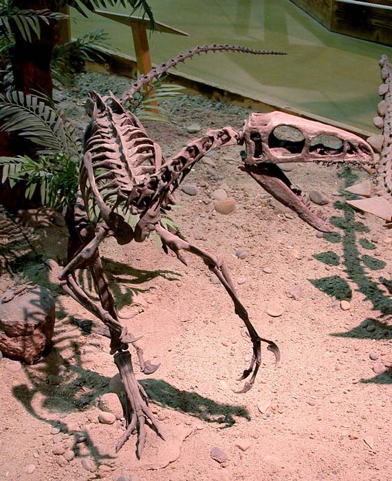
A coelurosaur, a subgroup of therapods who produced the archaeopteryx (a genus of bird-like dinosaurs) and modern birds. (Greg Goebel / CC BY-SA 2.0 )
Previous Finds that Support This Hypothesis
It was in China itself that early “feather” finds in an intermediary species helped first posit this theory. In the 1990s, a bunch of fossils were discovered that lacked wings, and had a panoply of plumage, fuzzy bristles, and fully articulated quills! Up until this point, paleontologists believed that feathers were unique to birds, but clearly there was an earlier history of feather evolution that originated before birds, as per this report in the Scientific American .
Interestingly, Professor Brusatte was also a co-author on a recent study published in Current Biology in 2014, that analyzed fossils from coelurosaurs, a subgroup of therapods who produced the archaeopteryx (a genus of bird-like dinosaurs) and modern birds. This study focused on small skeletal changes to show that there was no great leap, but instead involved small, gradual evolutionary steps over time.
“A bird didn’t just evolve from a T. rex overnight, but rather the classic features of birds evolved one by one; first bipedal locomotion, then feathers, then a wishbone, then more complex feathers that look like quill-pen feathers, then wings,” Brusatte said. “The end result is a relatively seamless transition between dinosaurs and birds, so much so that you can’t just draw an easy line between these two groups.”
It is clear that there is so much we don’t know but if anything the picture is becoming clearer on how dinosaurs evolved to become feather birds. Co-author on the current study, Xing Lida, sums up the find. She argues that the current hypothesis needs to be tested further with more embryo fossil finds, which are hard to come by, but worth the sweat and patience.
Top image: Representational image showing dinosaur embryo in an egg. Source: KtD / Adobe Stock