- Precision functional MRI mapping reveals distinct connectivity patterns for depression associated with traumatic brain injury Science
- Redefining Depression: TBI Affective Syndrome Discovered Neuroscience News
- Depression after a brain injury is a distinct condition, study finds. That could change how it’s treated. AOL
- Why depression after traumatic brain injury is distinct — and less likely to respond to standard treatment STAT
- Abnormal Brain Folding a Biomarker for Major Depressive Disorder Neuroscience News
- View Full Coverage on Google News
Tag Archives: connectivity
Essential Signaling Pathway for Neuronal Connectivity During Brain Development Identified
Summary: Study reveals a signaling pathway that controls the formation of synapses between pyramidal neurons and inhibitory neurons expressing the parvalbumin protein.
Source: King’s College London
New research from the Institute of Psychiatry, Psychology & Neuroscience (IoPPN) at King’s College London has demonstrated that brain wiring requires the control of local protein synthesis at the level of specific synapse types.
In new research published in Science, a collaborative study between the Rico and Marín groups reported that the regulation of protein synthesis occurs in a highly specific manner, to the degree of the type of synapse involved.
The authors identified a signaling pathway controlling the formation of synapses between excitatory pyramidal cells and inhibitory interneurons expressing the protein parvalbumin.
This is the first study that demonstrates the presence of such specificity in the regulation of protein synthesis during brain wiring.
The cerebral cortex is the outer layer of the human brain’s largest part, the cerebrum. It is responsible for our most sophisticated and diverse behaviors through its control of motor and sensory functions. It is also one of the most complex biological systems, so understanding the mechanisms that control its development is a major scientific challenge.
There are two main types of neurons in the cerebral cortex: excitatory pyramidal cells and inhibitory interneurons. The interaction between each part is crucial for the normal function of the cerebral cortex. Inhibitory interneurons pace and synchronize the activity of excitatory neurons, thereby orchestrating their behavior.
Neurons in the cerebral cortex organize in networks wired by connections known as synapses. Like an electrical connection, synapses consist of pre- (power plug) and post-synaptic (socket) compartments. In the adult brain, protein synthesis occurs locally in both compartments to carry out the function of the neurons.
Controlling the synthesis of specific proteins, through chemical signaling, allows the brain to regulate the activities of individual synapses. How this regulation differs between two types of developing cerebral cortex neurons, however, was not fully understood.
“Exploring the molecular processes regulating the development of cortical connectivity is thrilling, especially when they end up being so specific. We identified a signaling pathway that controls protein synthesis in one of the most fundamental connections in the cerebral cortex, the synapses made by pyramidal cells on parvalbumin interneurons,” says Dr. Clémence Bernard, the first author of the study from King’s IoPPN.
Abnormal protein synthesis in synapses is a core mechanism underlying ASD. The mechanism identified in this paper reveals an interplay of proteins associated with neurodevelopmental disorders. This discovery supports the idea that the synapses made by excitatory pyramidal cells and the parvalbumin-positive interneurons might be particularly sensitive to dysregulation seen in developmental brain conditions such as ASD.
“It’s fascinating that many genes linked to ASD seem to be regulated by the same signaling pathway we have identified in this study,” says Professor Marín, one of the two senior authors of the study.
“This observation suggests that the connections between excitatory pyramidal cells and inhibitory interneurons expressing parvalbumin are a possible hot spot for multiple genetic risk factors in ASD,” says Professor Rico, co-senior author of the study.
About this neuroscience research news
Author: Press Office
Source: King’s College London
Contact: Press Office – King’s College London
Image: The image is credited to King’s College London
Original Research: Closed access.
“Cortical wiring by synapse type–specific control of local protein synthesis” by Clémence Bernard et al. Science
See also


Abstract
Cortical wiring by synapse type–specific control of local protein synthesis
Neurons use local protein synthesis to support their morphological complexity, which requires independent control across multiple subcellular compartments up to the level of individual synapses.
We identify a signaling pathway that regulates the local synthesis of proteins required to form excitatory synapses on parvalbumin-expressing (PV+) interneurons in the mouse cerebral cortex.
This process involves regulation of the TSC subunit 2 (Tsc2) by the Erb-B2 receptor tyrosine kinase 4 (ErbB4), which enables local control of messenger RNA {mRNA} translation in a cell type–specific and synapse type–specific manner.
Ribosome-associated mRNA profiling reveals a molecular program of synaptic proteins downstream of ErbB4 signaling required to form excitatory inputs on PV+ interneurons.
Thus, specific connections use local protein synthesis to control synapse formation in the nervous system.
Amygdala connectivity predicts ketamine treatment response among patients with anxious depression
A brain region known as the amygdala could play a key role in predicting symptom improvement following ketamine therapy in patients with treatment-resistant anxious depression, according to new research published in the Journal of Affective Disorders.
“Since the antidepressant effects of ketamine in patients with anxious depression remain unclear, it is necessary to investigate the potential biomarkers predicting the antidepressant efficacy of ketamine in patients with anxious depression,” said study author Bin Zhang of the Affiliated Brain Hospital of Guangzhou Medical University.
“Previous studies have pointed out that functional connectivity differences in the amygdala are linked to depression improvement after ketamine treatment in depressed patients, but their role in anxious depression patients is uncertain. Therefore, we investigated the correlation between depression improvement after ketamine treatment and amygdala functional connectivity in anxious depression patients.”
For their study, the researchers examined neuroimaging data from 31 patients with anxious depression and 18 patients with non-anxious depression.
The researchers only included participants who had a diagnosis of major depression without comorbid psychotic symptoms, had a score greater than 17 on the Hamilton Depression Rating Scale, had previously failed to improve after at least two antidepressant treatments, had completed fMRI brain scans, and had undergone six ketamine infusions.
Among the patients with anxious depression, about 60% (20 patients) exhibited clinically significant reductions in depression symptoms following their sixth ketamine infusion. The remaining 11 patients with anxious depression were classified as non-responders.
The researchers found that, prior to the ketamine infusions, those who responded to the treatment tended to have greater functional connectivity between the left laterobasal amygdala and the left precuneus compared to non-responders. Additionally, the connectivity between the two brain regions was significantly reduced post-treatment among responders.
Patients with anxious depression also tended to have reduced connectivity between the right centriomedial amgydala and the right middle temporal gyrus compared to patients with non-anxious depression, which predicted treatment response.
“Corresponding to the crucial role of the amygdala in emotion regulation, especially in negative emotion, our study shown that the amygdala functional connectivity is associated with depression improvement to ketamine infusions in patients with anxious depression,” Zhang told PsyPost.
“The most surprising finding of the current study was that the baseline hyperconnectivity of the amygdala-precuneus found in the responders relative to the non-responders was significantly reduced on day 13 compared to baseline after six ketamine infusions. It may point to a potential neural underpinning by which ketamine exerts its antidepressant effect in patients with anxious depression.”
The results provide new insights into the mechanisms underlying ketamine’s antidepressant effects. But as with any study, the new research includes limitations. The researchers noted that their sample size was relatively small. Future research with larger samples should be conducted to validate the findings.
“Though the findings in our study may suggest that amygdala functional connectivity is a significant predictor of treatment response to ketamine infusions in patients with anxious depression, further validation is required,” Zhang said. “Moreover, further studies exploring the potential antidepressant mechanisms of ketamine may aid in the treatment of anxious depression patients.”
The study, “Functional connectivity differences in the amygdala are related to the antidepressant efficacy of ketamine in patients with anxious depression“, was authored by Shiqi Yuan, Xin Luo, Xiaoyu Chen, Mingqia Wang, Yiru Hu, Yanling Zhou, Yuping Ning, and Bin Zhang.
for(var key in aepc_pixel_args) args[key] = aepc_pixel_args[key];
return args; };
// Extend args if ( 'yes' === aepc_pixel.enable_advanced_events ) { aepc_pixel_args.userAgent = navigator.userAgent; aepc_pixel_args.language = navigator.language;
if ( document.referrer.indexOf( document.domain ) < 0 ) {
aepc_pixel_args.referrer = document.referrer;
}
}
!function(f,b,e,v,n,t,s){if(f.fbq)return;n=f.fbq=function(){n.callMethod?
n.callMethod.apply(n,arguments):n.queue.push(arguments)};if(!f._fbq)f._fbq=n;
n.push=n;n.loaded=!0;n.version='2.0';n.agent="dvpixelcaffeinewordpress";n.queue=[];t=b.createElement(e);t.async=!0;
t.src=v;s=b.getElementsByTagName(e)[0];s.parentNode.insertBefore(t,s)}(window,
document,'script','https://connect.facebook.net/en_US/fbevents.js');
fbq('init', aepc_pixel.pixel_id, aepc_pixel.user);
setTimeout( function() {
fbq('track', "PageView", aepc_pixel_args);
}, aepc_pixel.fire_delay * 1000 );
Read original article here
Scientists Discover Depression Treatment Increases Brain Connectivity
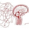
Representative map of the affected connections in the brain. The number of these connections increased after treatment. Credit: Jonathan Repple
Treatment for depression increases brain connectivity.
Most scientists believe that the structure of the adult brain is generally rigid and incapable of rapid changes. However, new research has now revealed that this is not true. In a new study, German scientists have shown that in-patient treatment for depression can lead to an increase in brain connectivity. Moreover, those individuals who respond well to this treatment show a greater increase in connectivity than those who don’t.
Presenting the work at the European College for Neuropsychopharmacology Congress in Vienna, lead researcher, Professor Jonathan Repple said:
“This means that the brain structure of patients with serious clinical depression is not as fixed as we thought, and we can improve brain structure within a short time frame, around 6 weeks. We found that if this treatment leads to an increase in brain connectivity, it is also effective in tackling depression symptoms. This gives hope to patients who believe nothing can change and they have to live with a disease forever, because it is “set in stone” in their brain.”


Graph showing the increase in the number of connections pre- and post-treatment for clinical depression. Credit: Jonathan Repple
Working at the University of Muenster in Germany, the scientists studied 109 patients with serious depression (Major Depressive Disorder) and compared them with 55 healthy controls. Participants’ brains were scanned using an MRI scanner that had been set up to identify which parts of the brain were communicating with other parts as a way of measuring the level of connections within the brain. The patients were then treated for depression, some with electroconvulsive therapy (ECT), some with psychological therapy or medication, and some with a combination of all therapies. After treatment, the study participants were then rescanned using MRI to measure the number of brain connections. They were also retested for symptoms of depression.
Professor Repple (now Professor of Predictive Psychiatry at the University of Frankfurt) said:
“We found that treatment for depression changed the infrastructure of the brain, which goes against previous expectations. Treated patients showed a greater number of connections than they had shown before treatment. Moreover, those who showed the most response to treatment developed a greater number of new connections than those who showed little response. A second scan showing that there are no time effects in healthy controls supports our findings that we see something that is related to the disease and more importantly the treatment of this disease.
“We found these changes took place over a period of only around 6 weeks, we were surprised at the speed of response. We don’t have an explanation as to how these changes take place, or why they should happen with such different forms of treatment.”
Commenting, Dr. Eric Ruhe, Rabdoud University Medical Center, Nijmegen, the Netherlands said:
“This is a very interesting and difficult-to-perform study in which the authors repeated MRI scans to reveal changes in structural connectivity over time in patients treated for depression.
“The results align very much with our current belief that the brain has much more flexibility in adaptation over (even short) time than was previously thought. Indeed a major idea of what treatment of depression (and other psychiatric illnesses) invoke is plastic changes over time. This has been proposed as a common mechanism for antidepressants, psychotherapy, and electroconvulsive therapy. However, the amount of research to elucidate what changes are necessary or specific for response to treatment or remission of depression is limited. Moreover, the next question is whether different treatments have the possibility to specifically change targeted brain networks or vice versa whether we can use the disturbances in brain-networks as measured in the present study to choose which therapy will be helpful.
“The fact that the observed changes over time could not be associated with a form of treatment is a pity, but as the authors themselves suggest a topic for further research. First these results should be replicated in independent samples which hopefully is going to happen soon. Second further elaboration on this approach would be daunting and should be supported firmly as this work might help to bridge the current gap between neuroscience and evidence-based patient care.”
This is an independent comment, Dr. Ruhe was not involved in this study.
This work was presented at the 35th European College of Neuropsychopharmacology annual conference, which took place in Vienna. The ECNP is Europe’s main organization working in applied neuroscience.
The Experience of Reward Increases Connectivity Between the Default Mode Network and Other Brain Regions
Summary: Study reveals how reward enhances connectivity between the ventral striatum and the default mode network, impacting behavior.
Source: Kessler Foundation
Researchers have reported findings that add to our knowledge of how human behavior may be shaped by the default mode network, a specific network of brain regions with both resting and task-related states.
The default mode network (DMN), which comprises the posterior medial cortex, medial prefrontal cortex, and lateral temporal-parietal regions, has been shown to be engaged in several task-related behaviors. Studies show that DMN activity increases during inward-directed thought and decreases during externally directed tasks requiring focused attention.
Despite evidence for a role for the DMN in shaping behavior, little is known about how task-related changes in the DMN influence connectivity with other brain regions. For example, while some observations indicate an indirect relationship between the DMN and the striatum, how the DMN and striatum interact during tasks remains unclear.
To further explore the functions of the DMN, Drs. Dobryakova and Smith applied a novel analysis to the reward task, using behavioral and neuroimaging data from 495 randomly selected individuals in the Human Connectome Project, an open access database of healthy participants.
The goals of this network-based psycho-physiological interaction analysis were twofold, according to Dr. Dobryakova, a senior research scientist in the Foundation’s Center for Traumatic Brain Injury Research.
“First, to test the effects of reward on connectivity between the DMN and the striatum; and second, whether such connectivity is associated with behavioral and personality characteristics relevant to reward processing,” she explained.
In line with other studies, during the task, they observed decreased activation of the DMN and relative increased activation of other networks.
“Most notably, we found that the experience of reward enhanced connectivity between the DMN and the ventral striatum,” Dr. Dobryakova reported, “an effect specific to the DMN. We were also surprised that the strength of this connectivity correlated with personality characteristics relating to openness,” she added.
Greater understanding of the workings of the healthy brain will influence future research and care for individuals with neuropsychiatric syndromes. “Improving our understanding of the interaction of the DMN with other brain networks has the potential to aid clinical research into better treatments for common syndromes such as depression, substance abuse, and schizophrenia,” Dr. Dobryakova concluded.
Funding: This research was supported by grants from the National Institutes of Health grants R21-MH113917 (DVS), R03-DA046733 (DVS), RF1-AG067011 (DVS), R01-NS121107 (ED).
About this reward and behavior neuroscience research news
Author: Carolann Murphy
Source: Kessler Foundation
Contact: Carolann Murphy – Kessler Foundation
Image: The image is in the public domain
Original Research: Open access.
“Reward enhances connectivity between the ventral striatum and the default mode network” by Ekaterina Dobryakova et al. NeuroImage
Abstract
See also
Reward enhances connectivity between the ventral striatum and the default mode network
The default mode network (DMN) has been theorized to participate in a range of social, cognitive, and affective functions. Yet, previous accounts do not consider how the DMN contributes to other brain regions depending on psychological context, thus rendering our understanding of DMN function incomplete.
We addressed this gap by applying a novel network-based psychophysiological interaction (nPPI) analysis to the reward task within the Human Connectome Project.
We first focused on the task-evoked responses of the DMN and other networks involving the prefrontal cortex, including the executive control network (salience network) and the left and right frontoparietal networks.
Consistent with a host of prior studies, the DMN exhibited a relative decrease in activation during the task, while the other networks exhibited a relative increase during the task. Next, we used nPPI analyses to assess whether these networks exhibit task-dependent changes in connectivity with other brain regions.
Strikingly, we found that the experience of reward enhances task-dependent connectivity between the DMN and the ventral striatum, an effect that was specific to the DMN. Surprisingly, the strength of DMN-VS connectivity was correlated with personality characteristics relating to openness.
Taken together, these results advance models of DMN by demonstrating how it contributes to other brain systems during task performance and how those contributions relate to individual differences.
Connectivity of Language Areas Unique in the Human Brain
Summary: Researchers shed new light on how the human brain evolved to be language-ready. Compared to the brains of chimps, the patterns of connections of language areas in the human brain expanded more than was previously thought.
Source: Radboud University
Neuroscientists have gained new insight into how our brain evolved into a language-ready brain. Compared to chimpanzee brains, the pattern of connections of language areas in our brain has expanded more than previously thought.
The researchers at Radboud University and University of Oxford publish their findings in PNAS on July 4.
“At first glance, the brains of humans and chimpanzees look very much alike. The perplexing difference between them and us is that we humans communicate using language, whereas non-human primates do not”, says co-first author Joanna Sierpowska.
Understanding what in the brain could have enabled this unique ability has inspired researchers for years. However, up to now, their attention was mainly drawn towards a particular nerve tract connecting frontal and temporal lobes called arcuate fasciculus, which besides showing significant differences between species, is well-known to be involved in language function.
”We wanted to shift our focus towards the connectivity of two cortical areas located in the temporal lobe, which are equally important for our ability to use language”, says Sierpowska.
To study the differences between the human and chimpanzee brain, the researchers used scans of 50 human brains and 29 chimpanzee brains scanned in a similar way as humans, but under well-controlled anesthesia and as part of their routine veterinary check-ups.
More specifically, they used a technique called diffusion-weighted imaging (DWI), which images white matter, the nerve pathways that connect brain areas.
Using these images, they explored the connectivity of two language-related brain hubs (the anterior and posterior middle areas of the temporal lobe), comparing them between the species.
“In humans, both of these areas are considered crucial for learning, using and understanding language and harbor numerous white matter pathways”, says Sierpowska.
“It is also known that damage to these brain areas has detrimental consequences for language function. However, until now, the question of whether their pattern of connections is unique to humans remained unanswered.”
See also
The researchers found that while the connectivity of the posterior middle temporal areas in chimpanzees is confined mainly to the temporal lobe, in humans a new connection towards the frontal and parietal lobes emerged using the arcuate fasciculus as an anatomical avenue. In fact, changes to both human language areas include a suite of expansions to connectivity within the temporal lobes.


“The results of our study imply that the arcuate fasciculus surely is not the only driver of evolutionary changes preparing the brain for a full-fledged language capacity”, says co-author Vitoria Piai.
“Our findings are purely anatomical, so it is hard to say anything about brain function in this context”, says Piai.
“But the fact that this pattern of connections is so unique for us humans suggests that it may be a crucial aspect of brain organization enabling our distinctive language abilities.”
About this language and evolutionary neuroscience research news
Author: Harriette Koop
Source: Radboud University
Contact: Harriette Koop – Radboud University
Image: The image is in the public domain
Original Research: The findings will appear in PNAS
Synaptic Connectivity of a Novel Cell Population in the Striatum
Summary: Researchers characterize a novel neural population within the striatum that appears to be responsible for the interplay between acetylcholine and GABA.
Source: Karolinska Institute
A new study from the Department of Neuroscience at Karolinska Institutet characterizes a novel neuronal population in the basal ganglia, responsible for the interaction between two types of neurotransmitters, GABA and acetylcholine.
The study was recently published in Cell Reports.
The striatum is the main input structure of the basal ganglia, a brain region involved in a variety of sensorimotor functions and reinforcement learning. 99% of striatal neurons are inhibitory GABAergic cells, and the only exception is the population of cholinergic interneurons.
“In previous studies we have showed the interactions between cholinergic interneurons and the dopamine system, and here we focused on the interactions between the cholinergic and GABAergic systems in the striatum,” explains Gilad Silberberg, Professor at the Department of Neuroscience, and main author of the study.
The striatum is strongly modulated by acetylcholine and early treatment for Parkinson’s disease was based on the cholinergic system. Cholinergic interneurons have been shown to change their activity in Parkinson’s disease, Huntington’s disease and in various forms of dyskinesia, all of which are disorders related to striatal function.
“Here we wanted to study how the cholinergic activity shapes striatal activity via nicotine receptors, a specific receptor of acetylcholine,” says Anna Tokarska, Ph.D. student in the Silberberg laboratory and first author of the study.
“To do that, we used transgenic mice, marking the striatal interneurons expressing these nicotinic receptors through the Chrna2 gene. We could then use various methods including patch-clamp and optogenetics, to characterize these neurons and their synaptic connectivity in the striatum,” she continues.
The striatal Chrna2 interneuron population was very diverse, including at least three main subpopulations with distinct anatomical and electrical properties. One population was of particular interest, showing novel characteristics including strong response to acetylcholine.
Future steps in this line of research will be to study this population in further detail, including its involvement in striatal function and dysfunction.
About this neuroscience research news
Author: Press Office
Source: Karolinska Institute
Contact: Press Office – Karolinska Institute
Image: The image is credited to Anna Tokarska, Gilad Silberberg
Original Research: Open access.
“GABAergic interneurons expressing the α2 nicotinic receptor subunit are functionally integrated in the striatal microcircuit” by Anna Tokarska et al. Cell Reports
Abstract
GABAergic interneurons expressing the α2 nicotinic receptor subunit are functionally integrated in the striatal microcircuit
See also

Highlights
- Triple whole-cell recordings are used to study striatal interneurons in Chrna2-Cre mice
- Unlike in other brain regions, most striatal Chrna2-interneurons express parvalbumin
- Three distinct subtypes of striatal Chrna2-interneurons are defined
- Their synaptic connectivity is mapped using optogenetics and patch-clamp recordings
Summary
The interactions between the striatal cholinergic and GABAergic systems are crucial in shaping reward-related behavior and reinforcement learning; however, the synaptic pathways mediating them are largely unknown.
Here, we use Chrna2-Cre mice to characterize striatal interneurons (INs) expressing the nicotinic α2 receptor subunit.
Using triple patch-clamp recordings combined with optogenetic stimulations, we characterize the electrophysiological, morphological, and synaptic properties of striatal Chrna2-INs.
Striatal Chrna2-INs have diverse electrophysiological properties, distinct from their counterparts in other brain regions, including the hippocampus and neocortex.
Unlike in other regions, most striatal Chrna2-INs are fast-spiking INs expressing parvalbumin. Striatal Chrna2-INs are intricately integrated in the striatal microcircuit, forming inhibitory synaptic connections with striatal projection neurons and INs, including other Chrna2-INs. They receive excitatory inputs from primary motor cortex mediated by both AMPA and NMDA receptors.
A subpopulation of Chrna2-INs responds to nicotinic input, suggesting reciprocal interactions between this GABAergic interneuron population and striatal cholinergic synapses.
Heightened dream recall ability linked to increased creativity and functional brain connectivity
People who can frequently recall their dreams tend to be more creative and exhibit increased functional connectivity in a key brain network, according to new research published in the journal Nature and Science of Sleep. The findings provide new insights into the neurophysiological correlates of dreaming.
“I think that dreaming is one of the last frontiers of human cognition — a terra incognita of the mind if you will,” said study author Raphael Vallat, a postdoctoral researcher at the Center for Human Sleep Science at the University of California, Berkeley. “Although we all spend a significant amount of our lives dreaming, there are still so many basic research questions related to dreams that are unanswered, which obviously makes it such a fascinating topic to study!
“In this and previous studies, we address one of these fundamental research questions: why do some people recall their dreams every day while others almost never seem to recall a dream?”
For his new study, Vallat and his colleagues used brain imaging techniques to examine whether neurophysiological differences exist between individuals who frequently recall their dreams and those who do not.
The study included 55 healthy participants (ages 19–29) with normal sleep characteristics and body mass index. Twenty-eight participants were high dream recallers (able to recall about 6 dreams per week on average), while 27 participants were low dream recallers (recalling less than one dream per week on average). The two groups did not significantly differ in age, habitual sleep duration, or education.
Participants arrived at the sleep lab at Le Vinatier Hospital the night before their scanning session and completed self-reported assessments of personality, anxiety, and sleep quality. They also completed the Wechsler Memory Scale (used to measure immediate and delayed memory performance), the Guildford Uses Task (used to measure creative ability), and a digit span task (used to measure working memory’s number storage capacity). After staying at the lab overnight, the participants underwent three functional magnetic resonance imaging scans to measure resting-state brain activity.
The researchers found that high dream recallers and low dream recallers had similar personalities, levels of anxiety, sleep quality, and memory abilities. However, high dream recallers scored significantly higher on the Guildford Uses Task than low dream recallers, indicating that they had greater creative abilities.
Vallat and his colleagues also observed increased functional connectivity within the default mode network in high dream recallers compared to low dream recallers. The brain network “is known to be active during day-dreaming, mind-wandering (e.g. getting lost in your thoughts), and has been further suggested to promote creativity and dreaming,” Vallat explained. The increased connectivity was specifically found between the medial prefrontal cortex and the temporo-parietal junction, in line with clinical reports that have shown lesions to these brain regions result in a cessation of dream recall.
“In simpler words, high dream recallers have superior creative abilities, as well as a different brain functional organization, as demonstrated by this study and previous studies from our lab,” Vallat told PsyPost. “It remains an open question whether there is a causal relationship between dream recall, creative thinking, and brain ‘wiring’, and if so, what is the direction of that relationship (the chicken or egg problem). Does increased dreaming promote creative thinking and ultimately lead to changes in brain function? Or does an innate higher functional connectivity of the default mode network in these individuals promote their dream recall and creative abilities?”
An experimental methodology could help to untangle the causal relationships. “A next step of this study could be to take a group of non-dreamers, increase their dream recall abilities over time using some validated methods (the most known of which is to simply write down their dreams every morning as they wake up, the conscious effort of remembering their dreams eventually leading to a better recall of dreams), and assess their creativity and brain function before and after the manipulation,” Vallat explained.
But the study, like all research, includes some limitations. “Like most functional magnetic resonance imaging (fMRI) studies, we have used a fairly small sample size, which limits the generalizability of our findings (i.e. do these findings hold for a larger and more diverse population?),” Vallat said.
The study also only examined one type of creativity. In the Guildford Uses Task, participants are given two minutes to list as many alternative uses as possible for an everyday object. The total number of responses and the number of rare uses are used to measure a type of creative ability known as divergent thinking. “Creativity is an umbrella term that encompasses several concepts (e.g. convergent vs divergent thinking, problem solving, gist extraction, etc). In this study, we have measured a single subdomain of creativity,” Vallat noted.
“Understanding differences in dream recall between individuals is just one angle through which we are trying to decipher this fascinating and mysterious phenomenon that is dreaming,” Vallat said. “Studying dreams is a nightmare (sorry for the pun!) because it is not directly observable: we do not know exactly when dreaming happens during sleep, and we must therefore rely on waking up the sleeper to ask whether they were dreaming or not prior to awakening. Even then, this is imperfect because if they do not report any dreams, we cannot know for sure whether they were not dreaming or were in fact dreaming but immediately forgot their dream(s) upon waking.”
The study, “High Dream Recall Frequency is Associated with Increased Creativity and Default Mode Network Connectivity“, was authored by Raphael Vallat, Başak Türker, Alain Nicolas, and Perrine Ruby.
for(var key in aepc_pixel_args) args[key] = aepc_pixel_args[key];
return args; };
// Extend args if ( 'yes' === aepc_pixel.enable_advanced_events ) { aepc_pixel_args.userAgent = navigator.userAgent; aepc_pixel_args.language = navigator.language;
if ( document.referrer.indexOf( document.domain ) < 0 ) {
aepc_pixel_args.referrer = document.referrer;
}
}
!function(f,b,e,v,n,t,s){if(f.fbq)return;n=f.fbq=function(){n.callMethod?
n.callMethod.apply(n,arguments):n.queue.push(arguments)};if(!f._fbq)f._fbq=n;
n.push=n;n.loaded=!0;n.version='2.0';n.agent="dvpixelcaffeinewordpress";n.queue=[];t=b.createElement(e);t.async=!0;
t.src=v;s=b.getElementsByTagName(e)[0];s.parentNode.insertBefore(t,s)}(window,
document,'script','https://connect.facebook.net/en_US/fbevents.js');
fbq('init', aepc_pixel.pixel_id, aepc_pixel.user);
setTimeout( function() {
fbq('track', "PageView", aepc_pixel_args);
}, aepc_pixel.fire_delay * 1000 );
Read original article here
Google Pixel Watch battery and connectivity details emerge
We’ve seen quite a bit of the Google Pixel Watch over the past couple of weeks. First, it was a Pixel Watch render, then live photos of the device. Although its appearance may no longer be a mystery, there are still a lot of unanswered questions about the device. If a source is to be believed, new information has surfaced, giving us new details about the Google Pixel Watch.
According to the folks at 9to5Google, the unannounced Wear OS device will pack a 300 mAh battery. This is great to know, but there are just too many factors involved that prevent us from knowing what really counts, which is how long the Pixel Watch will last. But, you have to give it to them for trying to figure it out. The site extensively compared the Pixel Watch’s reported 300 mAh battery and found similarities with a few models, namely the Samsung Galaxy Watch 4, the Fossil Gen 6, and Skagen’s Faster Gen 6.
Utilizing the data, they speculated that the Pixel Watch could last anywhere from 20 to 48 hours. This is, of course, quite a large spread. But again, it will be hard to know without getting all of the details and even then, this is a device that could get better with time. Expanding its capabilities through software optimizations and updates. In addition to the battery, the source also confirmed that there would be cellular connectivity.
While it is unknown when the Google Pixel Watch will be announced, many speculate that it could arrive during the Google I/O 2022 developer conference. The conference, which takes place on May 11 and May 12 could play host to Google’s new wearable. Hopefully, the smartwatch will emerge during the event, priced to compete. Luckily, we won’t have to wait long as the event is only a couple of weeks away.
Source: 9to5Google
Rivian announces membership plan with complimentary charging and LTE connectivity
With R1T trucks rolling off the assembly line at its factory in Normal, Illinois, Rivian continues to prepare for the official debut of its first EVs later this month. On Thursday, the automaker introduced a membership program that will grant Rivian owners access to complimentary charging at its soon-to-be-built Adventure Network and Waypoints chargers. It also pledged to match every mile Rivian Membership customers drive with energy from renewable resources such as wind and solar, as well as offer unlimited access to 4G LTE connectivity.
Additionally, the service includes Rivian off-Roadside Assistance, additional coverage that will see the company send a recovery vehicle to you if you get stuck out on the trail or need an emergency battery recharge. The company also promised to add additional perks in the future, including new drive modes, community meetups and in-cabin content. Each new Rivian vehicle will come with 12 months of free access to the service. After that, you’ll need to pay to continue enjoying the perks of the membership. The company hasn’t said how much it plans to charge for the service, so we’ve reached out to it for more information.
All products recommended by Engadget are selected by our editorial team, independent of our parent company. Some of our stories include affiliate links. If you buy something through one of these links, we may earn an affiliate commission.