- Traumatic memories are represented differently than sad memories in the brains of people with PTSD, research shows Medical Xpress
- Brain Study Suggests Traumatic Memories Are Processed as Present Experience The New York Times
- Brain Activity in PTSD: Trauma Memories Differ from Sad Ones Neuroscience News
- Neural patterns unravel distinctions between traumatic and sad memories in individuals with PTSD News-Medical.Net
- PTSD patients’ brains work differently when recalling traumatic experiences Popular Science
- View Full Coverage on Google News
Tag Archives: brains
Jellyfish are not the ‘simple creatures’ once thought: New study may change an understanding of our own brains – Fox News
- Jellyfish are not the ‘simple creatures’ once thought: New study may change an understanding of our own brains Fox News
- Brainless Brilliance: Jellyfish Stun Scientists With Learning Skills SciTechDaily
- A species of jellyfish carrying one of the most deadly venoms in the world is capable of learning despite not having a brain, new research shows Yahoo! Voices
- Can Cells Learn? Can Molecules Communicate? What We Are Learning… Walter Bradley Center for Natural and Artificial Intelligence
- A species of jellyfish carrying one of the most deadly venoms in the world is capable of learning despite not Business Insider India
- View Full Coverage on Google News
Country-level gender inequality is associated with structural differences in the brains of women and men | Proceedings of the National Academy of Sciences – pnas.org
- Country-level gender inequality is associated with structural differences in the brains of women and men | Proceedings of the National Academy of Sciences pnas.org
- Gender discrimination may be making parts of the female brain thinner Medical Xpress
- Stress caused by gender inequality is damaging women’s brains, study finds The Independent
- Experiencing Gender Discrimination May Make Parts Of Women’s Brains Thinner IFLScience
- Gender inequality ‘shrinks women’s brains’, Oxford University study finds The Telegraph
- View Full Coverage on Google News
Some People’s Brains Are Wrinklier Than Others, And Now We Know Why : ScienceAlert
The folds of the human brain are instantly recognizable. Snaking ridges and deep furrows give the squishy tissue inside our heads structure and the appearance of a wrinkly walnut.
Into peaks called gyri and fissures called sulci, the outermost layer of brain tissue is folded so that reams of it can be squeezed into the skull, and it’s here, on the brain’s wrinkly surface, that memory, thinking, learning, and reasoning all happens.
This folding, or gyrification, is crucial for proper brain function and circuitry – and is said to be why humans have greater cognitive abilities than apes and elephants, whose brains have some folds, and rats and mice, whose smoothed-surfaced brains have none.
Now, a team of scientists has discovered why some people have more brain folds than others, in a condition that affects normal brain development called polymicrogyria (PMG).
In polymicrogyria, too many gyri are piled on top of each other, resulting in an abnormally thick cortex and leading to a broad spectrum of problems such as neurodevelopmental delay, intellectual disability, speech difficulties, and epileptic seizures.
“Until recently, most hospitals treating patients with this condition did not test for genetic causes,” explains University of California San Diego (UCSD) neuroscientist Joseph Gleeson, one of the researchers behind the new study.
Polymicrogyria comes in many forms, with localized or widespread cortical thickening detectable on brain scans.
Mutations in 30 genes and counting have been associated with the condition. But how any of those genetic errors, alone or in tandem, result in the overfolded brain tissue remains unclear. Many cases of PMG also lack an identifiable genetic cause.
It’s thought to have something to do with the tardy migration of cortical brain cells in early development that leads to a disordered cortex. The cortex is the outermost layer of the brain’s two-lobed cerebrum, a thin sheet of gray matter composed of billions of cells.
To investigate further, Gleeson collaborated with researchers at the Human Genetics and Genome Research Institute in Cairo to tap into a database of nearly 10,000 families from the Middle East affected by some form of pediatric brain disease.
They found four families with a nearly identical form of PMG, all harboring mutations in one gene. That gene encodes a protein that clings to the surface of cells, with the imaginative name of transmembrane protein 161B (TMEM161B). But no one knew what it did.
Gleeson and colleagues showed in subsequent experiments that TMEM161B is found in most fetal brain cell types: in progenitor cells that grow into specialized neurons, in mature neurons that excite or inhibit their neighbors, and in glial cells that support and protect neurons in various ways.
However, TMEM161B is from a family of proteins that first appeared, evolutionarily speaking, in sponges – which have no brain.
This puzzled Gleeson and fellow UCSD neuroscientist Lu Wang who wondered if the protein might indirectly affect cortical folding by meddling with some basic cellular properties that give shape to complex tissues.
“Once we identified TMEM161B as the cause, we set out to understand how excessive folding occurs,” says Wang, the study’s lead author.
Using stem cells derived from patient skin samples, the researchers generated organoids, tiny tissue replicas that self-organize in plastic dishes the way bodily tissues and organs do. But the organoids made from patient cells were highly disorganized and showed disrupted radial glial fibers.
In the developing brain, these progenitor cells – which give rise to neurons and glia – usually position themselves at the apex of the cortex and extend radially downwards towards the bottom layer of cortical tissue. This creates a scaffolding system that supports the migration of other newly formed cells as the cortex expands.
But without TMEM161B, radial glial fibers in the organoids had lost sense of which way to orient themselves. Further experiments also showed that the cells’ internal cytoskeleton was a mess.
So it seems that without their own internal scaffold, radial glial fibers cannot be the scaffold other cells need to find their way into position in the developing brain.
While this discovery is a promising step forward, giving us clues to how the condition unfolds, it may only be relevant to a small or as yet unknown fraction of PMG cases.
Much more research is needed to flesh out our understanding of how many people with PMG are affected by mutations in TMEM161B – but now researchers know what to look for, they can trawl other datasets looking for more cases.
“We hope that physicians and scientists can expand upon our results to improve diagnosis and care of patients with brain disease,” says Gleeson. That’s a long road but a hopeful one.
The study has been published in PNAS.
The Brain’s Ability to Perceive Space Expands Like the Universe
Summary: Time spent in a novel environment causes neural representations to grow in a surprising way.
Source: Salk Institute
Young children sometimes believe that the moon is following them, or that they can reach out and touch it. It appears to be much closer than is proportional to its true distance. As we move about our daily lives, we tend to think that we navigate space in a linear way.
But Salk scientists have discovered that time spent exploring an environment causes neural representations to grow in surprising ways.
The findings, published in Nature Neuroscience on December 29, 2022, show that neurons in the hippocampus essential for spatial navigation, memory, and planning represent space in a manner that conforms to a nonlinear hyperbolic geometry—a three-dimensional expanse that grows outward exponentially. (In other words, it’s shaped like the interior of an expanding hourglass.)
The researchers also found that the size of that space grows with time spent in a place. And the size is increasing in a logarithmic fashion that matches the maximal possible increase in information being processed by the brain.
This discovery provides valuable methods for analyzing data on neurocognitive disorders involving learning and memory, such as Alzheimer’s disease.
“Our study demonstrates that the brain does not always act in a linear manner. Instead, neural networks function along an expanding curve, which can be analyzed and understood using hyperbolic geometry and information theory,” says Salk Professor Tatyana Sharpee, holder of the Edwin K. Hunter Chair, who led the study.
“It is exciting to see that neural responses in this area of the brain formed a map that expanded with experience based on the amount of time devoted in a given place. The effect even held for miniscule deviations in time when animal ran more slowly or faster through the environment.”
Sharpee’s lab uses advanced computational approaches to better understand how the brain works. They recently pioneered the use of hyperbolic geometry to better understand biological signals like smell molecules, as well as the perception of smell.
In the current study, the scientists found that hyperbolic geometry guides neural responses as well. Hyperbolic maps of sensory molecules and events are perceived with hyperbolic neural maps.
The space representations dynamically expanded in correlation with the amount of time the rat spent exploring each environment. And, when a rat moved more slowly through an environment, it gained more information about the space, which caused the neural representations to grow even more.
“The findings provide a novel perspective on how neural representations can be altered with experience,” says Huanqiu Zhang, a graduate student in Sharpee’s lab.
“The geometric principles identified in our study can also guide future endeavors in understanding neural activity in various brain systems.”
“You would think that hyperbolic geometry only applies on a cosmic scale, but that is not true,” says Sharpee.
“Our brains work much slower than the speed of light, which could be a reason that hyperbolic effects are observed on graspable spaces instead of astronomical ones. Next, we would like to learn more about how these dynamic hyperbolic representations in the brain grow, interact, and communicate with one another.”
Other authors include P. Dylan Rich of Princeton University and Albert K. Lee of the Janelia Research Campus at the Howard Hughes Medical Institute.
See also


About this spatial perception research news
Author: Press Office
Source: Salk Institute
Contact: Press Office – Salk Institute
Image: The image is credited to Salk Institute
Original Research: Open access.
“Hippocampal spatial representations exhibit a hyperbolic geometry that expands with experience” by Huanqiu Zhang et al. Nature Neuroscience
Abstract
Hippocampal spatial representations exhibit a hyperbolic geometry that expands with experience
Daily experience suggests that we perceive distances near us linearly. However, the actual geometry of spatial representation in the brain is unknown.
Here we report that neurons in the CA1 region of rat hippocampus that mediate spatial perception represent space according to a non-linear hyperbolic geometry. This geometry uses an exponential scale and yields greater positional information than a linear scale.
We found that the size of the representation matches the optimal predictions for the number of CA1 neurons. The representations also dynamically expanded proportional to the logarithm of time that the animal spent exploring the environment, in correspondence with the maximal mutual information that can be received. The dynamic changes tracked even small variations due to changes in the running speed of the animal.
These results demonstrate how neural circuits achieve efficient representations using dynamic hyperbolic geometry.
Alzheimer’s Disease Researchers Study Gene Associated With the Brain’s Immune Cells
Summary: Reduction of the INPP5D gene variant found in the brain’s microglia could help to diminish the risk of late-onset Alzheimer’s disease.
Source: Indiana University
Indiana University School of Medicine researchers are studying how the reduction of a gene variant found in the brain’s immune cells could diminish the risk of late-onset Alzheimer’s disease.
The research team, led by Adrian Oblak, Ph.D., assistant professor of radiology and imaging sciences, and Peter Bor-Chian Lin, a Ph.D. candidate in the Medical Neuroscience Graduate Program at Stark Neurosciences Research Institute, recently published their findings in Alzheimer’s & Dementia.
They focused their investigation on INPP5D, a microglia-specific gene that has been shown to increase the risk for developing late-onset Alzheimer’s disease. Microglia are the brain’s immune cells and there are multiple microglial genes associated with neurodegeneration.
Oblak said the team’s previous data revealed that elevated levels of INPP5D in Alzheimer’s disease lab models resulted in increased plaque deposition. Knowing this, they aimed to understand how reducing expression of INPP5D might regulate disease pathogenesis.
Using models in the lab, the researchers reduced the expression of the gene by at least 50%—called haplodeficiency—rather than completely knocking out the expression of the gene to mimic the treatment of pharmacological inhibitors targeting INPP5D as therapeutic strategies.
“INPP5D deficiency increases amyloid uptake and plaque engagement in microglia,” Oblak said. “Furthermore, inhibiting the gene regulates microglial functions and mitigates amyloid pathology that are likely mediated by TREM2-SYK signaling pathway activation.”
The gene deficiency also led to the preservation of cognitive function in the lab models. By reducing the expression of the gene in the brain, it created a less neurotoxic environment and improved the movement of microglia—which act as the first line of defense against viruses, toxic materials and damaged neurons—to clear amyloid deposits and plaques.
“These findings suggest that mitigating the function of INPP5D can result in a protective response by diminishing disease risk and mitigating the effect of amyloid beta induced pathogenesis,” Lin said.
About this Alzheimer’s disease and genetics research news
Author: Press Office
Source: Indiana University
Contact: Press Office – Indiana University
Image: The image is in the public domain
Original Research: Open access.
“INPP5D deficiency attenuates amyloid pathology in a mouse model of Alzheimer’s disease” by Peter Bor‐Chian Lin et al. Alzheimer’s & Dementia
See also

Abstract
INPP5D deficiency attenuates amyloid pathology in a mouse model of Alzheimer’s disease
Introduction
Inositol polyphosphate-5-phosphatase (INPP5D) is a microglia-enriched lipid phosphatase in the central nervous system. A non-coding variant (rs35349669) in INPP5D increases the risk for Alzheimer’s disease (AD), and elevated INPP5D expression is associated with increased plaque deposition. INPP5D negatively regulates signaling via several microglial cell surface receptors, including triggering receptor expressed on myeloid cells 2 (TREM2); however, the impact of INPP5D inhibition on AD pathology remains unclear.
Methods
We used the 5xFAD mouse model of amyloidosis to assess how Inpp5d haplodeficiency regulates amyloid pathogenesis.
Results
Inpp5d haplodeficiency perturbs the microglial intracellular signaling pathways regulating the immune response, including phagocytosis and clearing of amyloid beta (Aβ). It is important to note that Inpp5d haploinsufficiency leads to the preservation of cognitive function. Spatial transcriptomic analysis revealed that pathways altered by Inpp5d haploinsufficiency are related to synaptic regulation and immune cell activation.
Conclusion
These data demonstrate that Inpp5d haplodeficiency enhances microglial functions by increasing plaque clearance and preserves cognitive abilities in 5xFAD mice. Inhibition of INPP5D is a potential therapeutic strategy for AD.
Scientists Grew Stem Cell ‘Mini Brains’ And Then The Brains Sort-of Developed Eyes : ScienceAlert
Mini brains grown in a lab from stem cells spontaneously developed rudimentary eye structures, scientists reported in a fascinating 2021 paper.
On tiny, human-derived brain organoids grown in dishes, two bilaterally symmetrical optic cups were seen to grow, mirroring the development of eye structures in human embryos. This incredible result could help us to better understand the process of eye differentiation and development, as well as eye diseases.
“Our work highlights the remarkable ability of brain organoids to generate primitive sensory structures that are light sensitive and harbor cell types similar to those found in the body,” said neuroscientist Jay Gopalakrishnan of University Hospital Düsseldorf in Germany in a 2021 statement.
“These organoids can help to study brain-eye interactions during embryo development, model congenital retinal disorders, and generate patient-specific retinal cell types for personalized drug testing and transplantation therapies.”
Brain organoids are not true brains, as you might be thinking of them. They are small, three-dimensional structures grown from induced pluripotent stem cells – cells harvested from adult humans and reverse-engineered into stem cells, that have the potential to grow into many different types of tissue.
In this case, these stem cells are coaxed to grow into blobs of brain tissue, without anything resembling thoughts, emotions, or consciousness. Such ‘mini brains’ are used for research purposes where using actual living brains would be impossible, or at the very least, ethically tricky – testing drug responses, for example, or observing cell development under certain adverse conditions.
This time, Gopalakrishnan and his colleagues were seeking to observe eye development.
In previous research, other scientists had used embryonic stem cells to grow optic cups, the structures that develop into almost the entire globe of the eye during embryonic development. And other research had developed optic cup-like structures from induced pluripotent stem cells.
Rather than grow these structures directly, Gopalakrishnan’s team wanted to see if they could be grown as an integrated part of brain organoids. This would add the benefit of seeing how the two types of tissue can grow together, rather than just growing optic structures in isolation.
“Eye development is a complex process, and understanding it could allow underpinning the molecular basis of early retinal diseases,” the researchers wrote in their paper.
“Thus, it is crucial to study optic vesicles that are the primordium of the eye whose proximal end is attached to the forebrain, essential for proper eye formation.”
Previous work in the development of organoids showed evidence of retinal cells, but these did not develop optic structures, so the team changed their protocols.
They didn’t attempt to force the development of purely neural cells at the early stages of neural differentiation and added retinol acetate to the culture medium as an aid to eye development.


Their carefully tended baby brains formed optic cups as early as 30 days into development, with the structures clearly visible at 50 days. This is consistent with the timing of eye development in the human embryo, which means these organoids could be useful for studying the intricacies of this process.
There are other implications, too. The optic cups contained different retinal cell types, which organized into neural networks that responded to light, and even contained lens and corneal tissue. Finally, the structures displayed retinal connectivity to regions of the brain tissue.
“In the mammalian brain, nerve fibers of retinal ganglion cells reach out to connect with their brain targets, an aspect that has never before been shown in an in vitro system,” Gopalakrishnan said.
And it’s reproducible. Of the 314 brain organoids the team grew, 73 percent developed optic cups. The team hopes to develop strategies for keeping these structures viable on longer time-scales for performing more in-depth research with huge potential, the researchers said.
“Optic vesicle-containing brain organoids displaying highly specialized neuronal cell types can be developed, paving the way to generate personalized organoids and retinal pigment epithelial sheets for transplantation,” they wrote in their paper.
“We believe that [these] are next-generation organoids helping to model retinopathies that emerge from early neurodevelopmental disorders.”
The research has been published in Cell Stem Cell.
A version of this article was first published in August 2021.
Stranded Dolphins Show Signs of Alzheimer’s Disease in Their Brains : ScienceAlert
Scientists have discovered markers of Alzheimer’s disease in the brains of three different species of dolphin found deceased, stranded onshore.
Evidence of mass cetacean strandings exists from before our own recorded history, yet why dolphins and whales beach themselves in groups is an enduring mystery.
While a direct link has been found between naval sonar and some beaked whales, and some individual animals washed up on shore have been clearly unwell, some with a belly full of plastic waste, most mass strandings provide little to no clues.
Toothed whales (Odontocetes) share a number of traits with humans, including (in at least five species that we know of) menopause. Their ability to live well beyond their reproductive years means they have the potential to be susceptible to late-onset diseases as well.
Alzheimer’s is the most common cause of disability in aging humans, gradually impairing memory, learning, and communication. Now it appears a similar affliction may impact our water-dwelling mammalian relatives too.
“I have always been interested in answering the question: do only humans get dementia?” says neurobiologist Frank Gunn-Moore from University of St Andrews in Scotland.
“Our findings answer this question as it shows potential dementia associated pathology is indeed not just seen in human patients.”
Leiden University biologist Marissa Vacher and colleagues examined the brains of 22 stranded dolphins to search for the biochemical markers present in humans with Alzheimer’s. These include amyloid-beta plaques, which while no longer thought to be a direct cause of the disease are still present in elevated numbers in those who have it; and clusters of tau proteins with hyperphosphorylation – when phosphate groups have been added to all possible binding sites on the protein molecule.
They found accumulations of amyloid-beta plaques and hyperphosphorylated tau in three dolphins, each from a different species: the long-finned pilot whale (Globicephala melas), the white-beaked dolphin (Lagenorhynchus albirostris) and the common bottlenose dolphin (Tursiops truncatus). These individuals also had signs of being elderly such as worn or lost teeth and an increase in the ratio of white to grey matter in brain tissues.
What’s more, the locations of brain lesions found in the dolphins matched with equivalent areas seen in humans with Alzheimer’s.
While it wasn’t possible for the researchers to verify an Alzheimer’s diagnosis, as they couldn’t test the deceased animals’ levels of cognitive impairment, there is no record of accumulations of both proteins in humans without the disease.
“We were fascinated to see brain changes in aged dolphins similar to those in human aging and Alzheimer’s disease,” says University of Edinburgh neuroscientist Tara Spires-Jones.
As dolphins are highly social animals, it’s possible they aid fellow pod members who begin to struggle with their brains. This means there’s a chance they’d survive for longer, allowing further progression of the disease than in solitary species, the researchers note.
Dolphin strandings are common in one of the species studied, G. melas, supporting the ‘sick-leader’ theory of this mysterious, fatal behavior.
“In humans, the first symptoms of AD-associated cognitive decline include confusion of time and place and a poor sense of direction,” Vacher and colleagues explain in their paper.
“If the leader of a pod of G. melas suffered from a similar neurodegenerative-related cognitive decline this could lead to disorientation resulting in leading the pod into shallow water and subsequent stranding.”
However, “whether these pathological changes contribute to these animals stranding is an interesting and important question for future work,” Spires-Jones concludes.
This research was published in the European Journal of Neuroscience.
Stranded Dolphins’ Brains Show Common Signs of Alzheimer’s Disease
Summary: Researchers found the brains of three species of stranded dolphins had classic biomarkers associated with human Alzheimer’s disease. The findings add weight to the “sick leader” theory, where a group of healthy dolphins finds themselves in dangerously shallow waters after following a group leader showing signs of confusion.
Source: University of Glasgow
The brains of three species of stranded dolphins show classic markers of human Alzheimer’s disease, according to the most extensive study into dementia in odontocetes (toothed whales).
The new pan-Scotland research, a collaboration between the University of Glasgow, the Universities of St Andrews and Edinburgh and the Moredun Research Institute, studied the brains of 22 odontocetes which had all been stranded in Scottish coastal waters.
The study, which is published in the European Journal of Neuroscience, included five different species—Risso’s dolphins, long-finned pilot whales, white-beaked dolphins, harbor porpoises and bottlenose dolphins—and found that four animals from different dolphin species had some of the brain changes associated with Alzheimer’s disease in humans.
The findings may provide a possible answer to unexplained live-stranding events in some odontocete species. Study authors confirm the results could support the “sick-leader” theory, whereby an otherwise healthy pod of animals find themselves in dangerously shallow waters after following a group leader who may have become confused or lost.
Whales, dolphins and porpoises are regularly stranded around the coasts of the U.K. They are often found stranded in groups, or pods, in shallow waters and sometimes on beaches. While some animals can be moved to safer, deeper waters by teams of experts, other animals are less lucky and perish as a result. The underlying causes of live stranding events are not always clear, and research is ongoing to gain better insights.
For this study researchers examined stranded animals for the presence of the brain pathology that are part of the hallmarks of Alzheimer’s disease, including the formation of amyloid-beta plaques, the accumulation of phospho-tau and gliosis (a change in cell numbers in response to central nervous system damage). The results reveal that the brains of all of the aged animals studied had amyloid-beta plaques.
Three animals in particular—each from a different odontocete species—had amyloid-beta plaques as well as a number of other dementia-related pathologies in their brains, showing that some odontocete species develop Alzheimer’s-like neuropathology. However, the study cannot confirm whether any of the animals would have suffered with the same cognitive deficits associated with clinical Alzheimer’s disease in humans.
Lead researcher, Dr. Mark Dagleish from the University of Glasgow, said, “These are significant findings that show, for the first time, that the brain pathology in stranded odontocetes is similar to the brains of humans affected by clinical Alzheimer’s disease.
“While it is tempting at this stage to speculate that the presence of these brain lesions in odontocetes indicates that they may also suffer with the cognitive deficits associated with human Alzheimer’s disease, more research must be done to better understand what is happening to these animals.”
Co-author, Professor Frank Gunn-Moore from the University of St Andrews, said, “I have always been interested in answering the question: do only humans get dementia? Our findings answer this question as it shows potential dementia associated pathology is indeed not just seen in human patients. This study is also a great example of both different research institutes, but also different branches of the Life Sciences working together.”
Professor Tara Spires-Jones, University of Edinburgh, said, “We were fascinated to see brain changes in aged dolphins similar to those in human aging and Alzheimer’s disease. Whether these pathological changes contribute to these animals stranding is an interesting and important question for future work.”
All animals in this research were studied after a stranding event. Marine Scotland and Defra fund post-mortem examinations, via the Scottish Marine Animal Stranding Scheme (SMASS), of cetaceans (including odontocetes), pinnipeds and marine turtles that strand and die in Scottish coastal waters.
About this animal neuroscience and Alzheimer’s disease research news
Author: Press Office
Source: University of Glasgow
Contact: Press Office – University of Glasgow
Image: The image is in the public domain
Original Research: Open access.
“Alzheimer’s disease‐like neuropathology in three species of oceanic dolphin” by Marissa C. Vacher et al. European Journal of Neuroscience
See also

Abstract
Alzheimer’s disease‐like neuropathology in three species of oceanic dolphin
Alzheimer’s disease (AD) is the most common neurodegenerative disease and the primary cause of disability and dependency among elderly humans worldwide. AD is thought to be a disease unique to humans although several other animals develop some aspects of AD-like pathology. Odontocetes (toothed whales) share traits with humans that suggest they may be susceptible to AD.
The brains of 22 stranded odontocetes of five different species were examined using immunohistochemistry to investigate the presence or absence of neuropathological hallmarks of AD: amyloid-beta plaques, phospho-tau accumulation and gliosis.
Immunohistochemistry revealed that all aged animals accumulated amyloid plaque pathology. In three animals of three different species of odontocete, there was co-occurrence of amyloid-beta plaques, intraneuronal accumulation of hyperphosphorylated tau, neuropil threads and neuritic plaques.
One animal showed well-developed neuropil threads, phospho-tau accumulation and neuritic plaques, but no amyloid plaques. Microglia and astrocytes were present as expected in all brain samples examined, but we observed differences in cell morphology and numbers between individual animals.
The simultaneous occurrence of amyloid-beta plaques and hyperphosphorylated tau pathology in the brains of odontocetes shows that these three species develop AD-like neuropathology spontaneously. The significance of this pathology with respect to the health and, ultimately, death of the animals remains to be determined.
However, it may contribute to the cause(s) of unexplained live-stranding in some odontocete species and supports the ‘sick-leader’ theory whereby healthy conspecifics in a pod mass strand due to high social cohesion.
Surprise Protector of Females’ Brains: Subcutaneous Fat
According to new research, subcutaneous fat, which is more common in females, is protective against brain inflammation.
Females’ propensity toward subcutaneous fat, which is fat stored under the skin, often in places like their hips, buttocks, and the backs of their arms, is protective against brain inflammation, at least until menopause. This is according to a new study by scientists at the Medical College of Georgia at Augusta University. It is important because brain inflammation can contribute to serious problems such as dementia and stroke.
Males of essentially any age, on the other hand, have a greater propensity to deposit fat around the major organs in their abdominal cavities. This is called visceral fat, or visceral adiposity adiposity, and is known to be far more inflammatory. And, before females reach menopause, males are considered at much higher risk for inflammation-related problems from heart attack to stroke.
“When people think about protection in women, their first thought is estrogen,” says Alexis M. Stranahan, PhD, neuroscientist in the Department of Neuroscience and Regenerative Medicine at the Medical College of Georgia at Augusta University. “But we need to get beyond the kind of simplistic idea that every sex difference involves hormone differences and hormone exposure. We need to really think more deeply about the underlying mechanisms for sex differences so that we can treat them and acknowledge the role that sex plays in different clinical outcomes.”
Diet and genetics are other likely factors that explain the differences broadly assigned to estrogen, says Stranahan, corresponding author of a study that was recently published in the American Diabetes Association journal Diabetes.
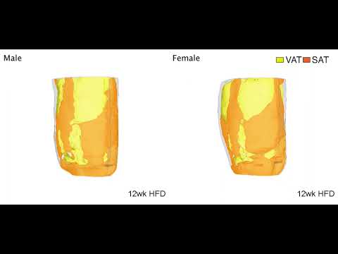
Watch a video of where males and female mice gain weight on a high-fat diet. While at some point females can have the same amount of visceral fat as males, there is still less inflammation. Credit: Alexis Stranahan, Medical College of Georgia
Stranahan acknowledges that the findings are potentially heretical and revolutionary and certainly surprising even to her. “We did these experiments to try and nail down, first of all, what happens first, the hormone perturbation, the inflammation, or the brain changes.”
To learn more about how the brain becomes inflamed, the researchers scrutinized increases in the amount and location of fat tissue as well as levels of sex hormones and brain inflammation in male and female mice at different time intervals as they grew fatter on a high-fat diet.
Since, much like with people, obese female mice tend to have more subcutaneous fat and less visceral fat than male mice, they reasoned that the distinctive fat patterns might be a key reason for the protection from inflammation the females enjoy before menopause.
In response to a high-fat diet, the investigators again found the distinctive patterns of fat distribution in males and females. They found no indicators of brain inflammation or insulin resistance, which also increase inflammation and can lead to diabetes, until after the female mice reached menopause. At about 48 weeks, menstruation stops and fat positioning on the females starts to shift somewhat, to become more like males.
They then compared the impact of the high-fat diet, which is known to increase inflammation body-wide, in mice of both sexes following surgery, similar to liposuction, to remove subcutaneous fat. They did nothing to directly interfere with normal estrogen levels, like removing the ovaries.
The subcutaneous fat loss increased brain inflammation in females without changing the levels of their estrogen and other sex hormones.
Bottom line: The females’ brain inflammation looked much more like the males’, including increased levels of classic inflammation promoters like the signaling proteins IL-1β and TNF alpha in the brain, Stranahan and her colleagues report.


Dr. Alexis Stranahan. Credit: Michael Holahan, Augusta University
“When we took subcutaneous fat out of the equation, all of a sudden the females’ brains start to exhibit inflammation the way that male brains do, and the females gained more visceral fat,” Stranahan says. “It kind of shunted everything toward that other storage location.” The transition occurred over about three months, which translates to several years in human time.
By comparison, it was only after menopause, that the females who did not have subcutaneous fat removed but did eat a high-fat diet, showed brain inflammation levels similar to the males, Stranahan says.
When subcutaneous fat was removed from mice on a low-fat diet at an early age, they developed a little more visceral fat and a little more inflammation in the fat. But Stranahan and her colleagues saw no evidence of inflammation in the brain.
One take-home lesson from the work: Don’t get liposuction and then eat a high-fat diet, Stranahan says. Another is: BMI, which simply divides weight by height and is commonly used to indicate overweight, obesity, and consequently increased risk of a myriad of diseases, is likely not a very meaningful tool, she says. An also easy and more accurate indicator of both metabolic risk and potentially brain health, is the also easy-to-calculate waist-to-hip ratio, she adds.
“We can’t just say obesity. We have to start talking about where the fat is. That is the critical element here,” Stranahan says.
She notes that the new study looked specifically in the hippocampus and hypothalamus of the brain. The hypothalamus controls metabolism and exhibits changes with inflammation from obesity that help control conditions that develop bodywide as a result. The hippocampus, a center of learning and memory, is regulated by signals associated with those pathologies but doesn’t control them, Stranahan notes. While these are good places to start such explorations, other regions of the brain could respond very differently, so she is already looking at the impact of loss of subcutaneous fat in others. Also, since her evidence indicates estrogen may not explain the protection females have, Stranahan wants to better define what does. One of her suspects is the clear chromosomal differences between the XX female and the XY male.
Stranahan has been studying the impact of obesity on the brain for several years and is among the first scientists to show that visceral fat promotes brain inflammation in obese male mice, and, conversely, transplanting subcutaneous fat reduces their brain inflammation. Females also have naturally higher levels of proteins that can tamp down inflammation. It’s been shown that in males, but not females, microglia, immune cells in the brain, are activated by a high-fat diet.
She notes that some consider the reason that females have higher stores of subcutaneous fat is to enable sufficient energy stores for reproduction, and she is not challenging the relationship. But many questions remain like how much fat is needed to maintain fertility versus the level that will affect your metabolism, Stranahan says.
Reference: ” Sex Differences in Adipose Tissue Distribution Determine Susceptibility to Neuroinflammation in Mice With Dietary Obesity” by Alexis M. Stranahan, De-Huang Guo, Masaki Yamamoto, Caterina M. Hernandez, Hesam Khodadadi, Babak Baban, Wenbo Zhi, Yun Lei, Xinyun Lu, Kehong Ding and Carlos M. Isales, 11 November 2022, Diabetes.
DOI: 10.2337/db22-0192
The research was supported by the National Institutes of Health (NIH).